Effects of structured physical activity in improving attention among school going children

Vishnupriya.R1, Srividya.G2, Kannan.D3

Corresponding Author:

1PhD Scholar, Annamalai University, Chidambaram, Tamil Nadu and Professor, JKKMMRF College of Physiotherapy, Komarapalayam, Namakkal, Tamil Nadu, India

Mail Id: vpvishnupriyaa114@gmail.com

Co-Authors:

2Research Guide, Department of PMR, Annamalai University, Chidambaram, Tamil Nadu, India

3Professor/Principal, JKKMMRF, College of Physiotherapy, Komarapalayam, Namakkal, Tamil Nadu, India.

 ABSTRACT

Background of the study: Attention is the behavioural and cognitive process of selectively concentrating on a discrete aspect of information. Difficulty in maintaining attention in the classroom is the main factor for lower grades. However, as an emerging outlook in physiotherapy, engaging the underachievers in structured physical activity will enhance a attention skill.  Consequently it will have a tremendous positive impact on children’s academic performance. This study aimed to identify the effect of structured physical activity in improving attention skill among school-going children.

Methodology: The study is an experimental study design and it was conducted among forty Children after obtaining ethical clearance and written consent from the school and parents of these children. All the children were selected based on the selection criteria. All the forty children were randomly allocated into two equal groups.  The Children in Group A underwent structured physical activity for 60 minutes per day for three days a week for a period of five months. The children in Group B experienced relaxed indoor activities for 60 minutes and were given three days a week for five months. The pre and post therapy assessment was done using Mindful attention awareness scale (MASS) questionnaire. The SPSS statistical package 26.0 was used to analyze and compare the collected data.

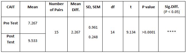
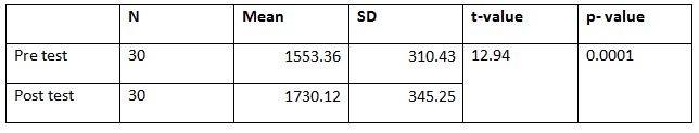
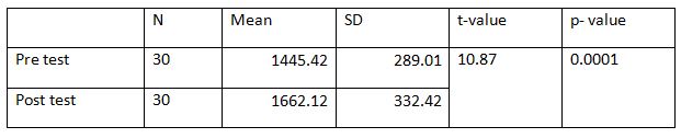
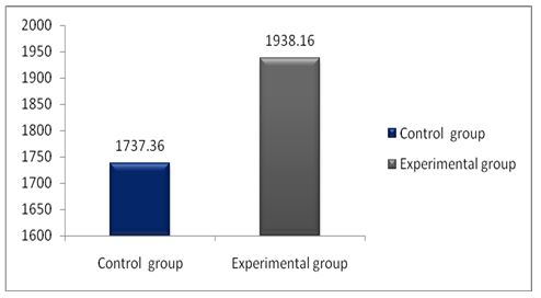
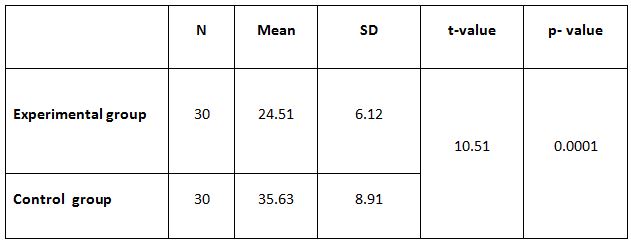
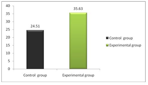
Result:  The study result revealed a mean score of 31.68 ± 0.85 for Group A and 14.80 ± 0.87 for Group B with the p-value of 0.0001 which is highly significant.

Conclusion: This study concludes that structured physical activity aids in improving the attention skill of school-going children which in turn improves the academic performance when compared with the children who underwent relaxed in door activities.

Keywords: Attention; MASS; Structured Physical Activity; Relaxed Indoor Activities.

Received on 24thDecember 2021, Revised on 14thFebruary 2022, Accepted on 25thFebruary 2022, DOI:10.36678/IJMAES.2022.V08I01.002

INTRODUCTION

Attention is the ability to choose and concentrate on relevant stimuli. It is the cognitive process that makes it possible   to position ourselves towards relevant stimuli and consequently respond to it. This cognitive ability is very important and is an essential function in students’ daily academic activities1.

In recent pandemic situation there has been a shift in the lifestyles of various age groups including children especially school going children as there was a break in the regular classroom education system of study2. Unlike children a few years ago children now a days are leading increasingly sedentary lifestyle that involves time spent on computer, Television and mobile gadgets.

Effective attention is what allows the children to screen out irrelevant stimulation in order to focus on the information that is important in the moment3. Difficulty with attention will lead to problems like not attending to a academic activity they miss details in instruction4. Children repeatedly make the same mistakes and unable to listen to all the information presented to them.

Prolonged sedentary behavior reduces the attention of the child and reduces the focus on the tasks5. Physical activity in early childhood helps preschoolers establish a healthy lifestyle 6. Various educational objectives propose the physical activity on contribution in mental acuity, skills, and strategies essential for navigating challenges faced across the life span7.

Physical activity (PA) is any bodily movement produced by the muscles which require energy expenditures systematically and safely8.It also includes the motor behaviors such as daily and leisure activities, and it is considered a determinant of life and for general health status 9.  Various studies identified that Physical activity facilitates the release of neurotrophic factors, which increases the blood flow to the cerebrum, thereby improving the supply of glucose and lipids to the brain11,12.

The Mindful attention awareness scale is a 15-item scale designed to assess dispositional mindfulness. This scale strongly evaluates the psychometric properties which have been validated. It is unique to measure the consciousness related to predictive of a variety of self-regulation and well-being constructs 13.

Various researches widely acknowledge the physical and mental health benefits of physical activity,14, but there are fewer studies on their effects on cognitive and academic performances and few works were reported on this topic but not on the school children 15, and there is much difference in the exercises prescribed by various researchers and Very few studies have been done in the area of physiotherapy on this perspective. So, this study is aimed to identify the effect of physical activity in improving attention among school-going children.

METHODOLOGY

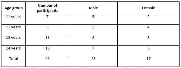
The study was initiated after obtaining ethical approval from the Institutional ethical committee, Annamalai University, Tamil Nadu, India. A pamphlet was created about physical activity and approached the private schools in and around Komarapalayam, Namakkal district, Tamil Nadu. The researcher met the principal and explained the study and around 15 schools were visited only ten school principals agreed to provide samples for the study. Written consent was obtained from the parent of the student who was included for the study prior to the beginning of the study. A meeting with the parent and the class teacher was made and explained the study. Only five schools were taken for this study, and 40 students were randomly selected from each school based on the inclusion criteria. The age group of the participants is 11 years to 14 years of both Genders.

The Children who had good attendance percentage and who were physically healthy with adaptive behavioral skills, were selected for the study. Children who are underachievers in their academic performance and children without any recent infections were also included for the study. Children with orthopedic or neurological disorders,   those with injury to the lower limbs, severely obese children, children with psychological disorders, children who are already in sports were excluded from the study.

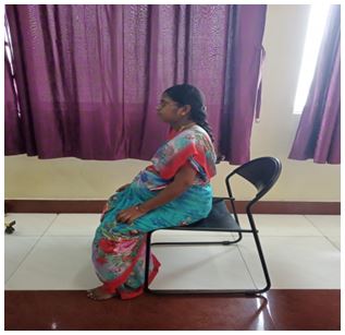
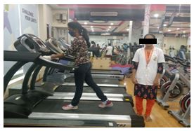
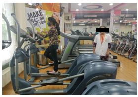
Forty-six children were selected for the study, and they all were divided randomly into two equal groups, twenty-three in each group. Group sampling was done by the computer-generated randomized method. The children in group A underwent structured physical activity for 60 minutes per day over a period of five month and three days per week. Structured physical activity (SPA) taught to the children includes walking with normal speed, walking with maximal speed, slow jogging, slow running, and skipping. Each exercise needs to be done for seven minutes16,17.

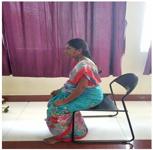
Group B underwent relaxed indoor activities (RIA) for 60 minutes per day over a period of 5 months and three days per week. Fifteen minutes were given for warming up exercises, and cool down, activities were also provided. The intensity of the exercises was set at 50%-70% of maximal heart rate 18.

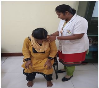
 A pre training data was collected using the MASS questionnaire. The post training data was collected by using the MASS Questionnaire at the end of the study (i.e., 5th month). The SPSS statistical package 26.0 was used to analyze and to compare the pre and post training on the impact of physical activity on the attention skill of school going children.  The impact of physical activity on the attention skills was analyzed before giving the structured physical activity again the data was collected after 5 months. It was statistically analyzed using SPSS statistical package.

The parametric test was used to determine the level of attention using MASS score. The scores obtained pre-test and post-test values were compared. The obtained p value of 0.05 showed the significant difference between pre and post. Due to illness,   three     children   were not able to attend the part of the study. As a result each groups both groups A and B from the study. As a result, each group had a total of 20 children.

RESULTS

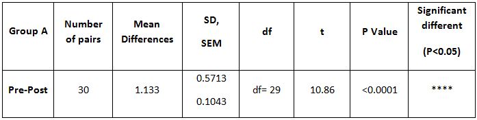
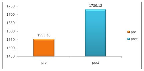
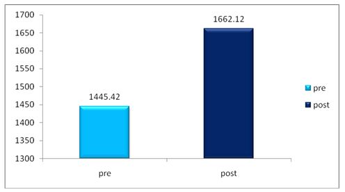
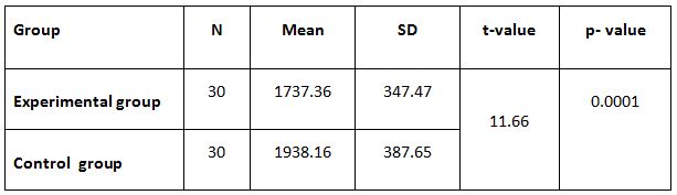
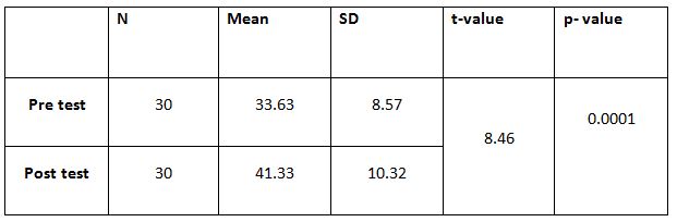
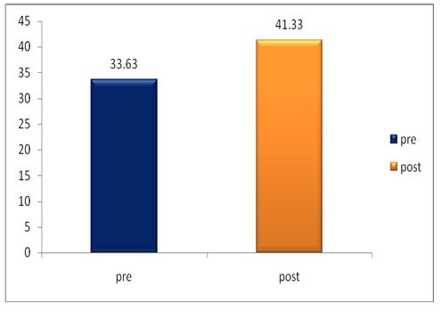
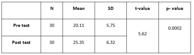
A parametric test is used to analyze the data collected from the children and the data were analyzed using SPSS 26.0. The with-in group analysis is shown in Table II, and the between-group analysis in Table III. The demographical variables are shown in Table I. All of the analysis in the study was done using a critical value of p= 0.05.
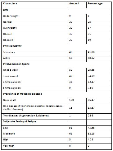
Table 1: Shows that out of 40 children, 13 children were 14 years and 11 children were 13 years and 16 children were in between 11 to 12 years there were more number of male children than female children.

 Food habits

Table 2: Demographic and Variable analysis
Table 3: Suggests the data within the groups of pre and post test values of Group A and Group B on analysis of MASS
* The probability of this result, assuming the null hypothesis, is less than .0001.
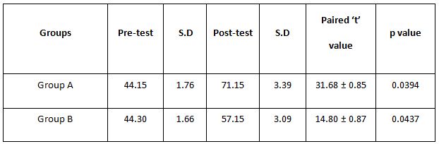
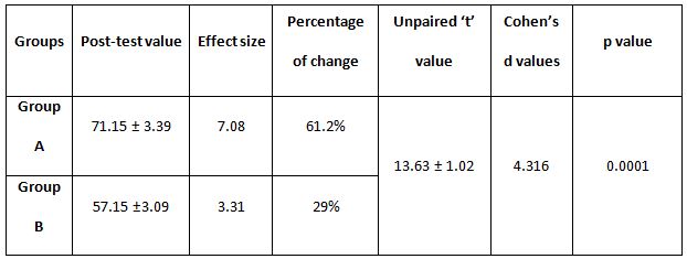
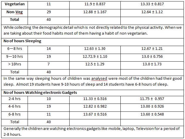
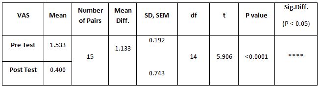
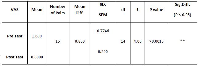
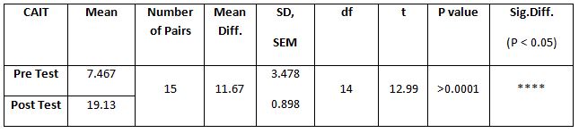
Table 4:  Showed the difference between Group A and Group B on analysis of MASS.

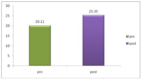
While comparing the pre-intervention and post-intervention data, there is a significant difference obtained between the groups. It was also noted there is a marked increase in attention skills of Group A students.

DISCUSSION

The purpose of the study was to identify the effects of structured physical activity in improving attention skill among school-going children. The problems of attention among the school going children are crucial in its contribution towards learning and academic performances19. School children have trouble having proper attention due to anxiety, frustration, and depression. A prolonged sedentary lifestyle reduces the child’s attention to academic instructions, concentration on the task engagements 20. So, it is mandatory to monitor these children and should give proper attention and training also needs to be given to them to overcome their difficulties 21

Physical activity (PA) is significant for a child to improve their fitness physically and mentally. It aids in controlling the various psychological symptoms and reduces the risk of developing multiple health issues22. Exercises improve children’s mental health by reducing anxiety, depression, and negative mood by improving self-esteem and cognitive functions23.

Physical activity plays an essential role in improving the child’s moods; the exercises increase circulation to the brain and influence the hypothalamic-pituitary-adrenal axis and reactive to stress24.

Many Studies also identified that acute bouts of aerobic exercises increase cerebral oxygenated blood flow and improve precuneus’ activity 25,26. The Physical activity also enhances Brain-Derived Neurotrophin Factors (BDNF) levels in the blood 27. This improves the activity of the brain and thereby improves the concentration, working memories, and visual-spatial abilities28,29

A few other studies also supported this study results; Tine and Butler have identified that 12 min single bout aerobic exercises improve attention and concentration in students with the age of 10-13 years30.

 A study conducted by Gallotta et al., 2012, identified that the school students involved in aerobic exercises in the physical education sessions showed an improved attention performance than those who didn’t participate31. Above study is supporting the present study.

In a study, Meta-analysis of   physical activity and cognitive performances in school-age children showed positive relationship. Several studies have suggested that participation in the PA has a positive relationship with academic performances.

All these findings have suggested that physical activity has a strong influence on academic performances and improvement in the attention of school children. It also plays a significant role in the development of cognitive health. 

This study showed a significant improvement in the attention skill of school children who underwent regular physical activity than those who were engaged in non-physical relaxed activity. The improvement in the relaxed activity group may be influence the participant in the research. There are considerable differences obtained within the groups. On comparing the groups, the group who underwent regular PA shows much significant improvement. 

This study has identified a few limitations, as selecting the participants and making parents accept to be involved in the study was a monumental task. Home activity or recreational sports activities are not controlled.

As the study duration was higher many students hesitated to participate in the study initially and later, which was liked by them, COVID-19 rules were followed, but the investigation was concluded by using telephone calls to individual parents and direct visits to each child by the researchers. 

Ethical Clearance: Ethical clearance has obtained from J.KK. Munirajah Medical Research Foundation, College of Physiotherapy, B. Komarapalayam, Tamil Nadu, to conduct this study with reference number: 001/jkk coptdated 04/06/2019.

Acknowledgment: The authors wish to thank the Principals of Schools, for allowing conducting the study and all the Children and the parents who were involved.

Conflict of Interest: There is no conflict of interest to conduct or publish this study.

Source of Funding:  This study is self funded.

CONCLUSION

This study concluded that structured physical activity is better in improving the attention of school-going children. When compare to the relaxed indoor activities.

REFERENCES

  1. Peretz C, Korczyn AD, Shatil E, Aharonson V, Birnboim S, Giladi N-Computer-Based, Personalized Cognitive Training versus Classical Computer games: A Randomized Double- Blind prospective Trial of cognitive stimulation-Neuroepide-miology 2011; 36: 91-9.
  2. Sedentary life style; Overview of updated evidence of potential health risks-jung ha park, Ji hyun moon and Yun hwan Oh. 2020 Nov; 41(6): 365–373.
  3. Plebanek, D. J., & Sloutsky, V. M. Costs of Selective Attention: When Children Notice What Adults Miss. Psychological science, 2017; 28(6), 723–732.
  4. Cotman CW, Berchtold NC, Christie L-A. Exercise builds brain health: key roles of growth factor cascades and inflammation. Trends Neurosci 2007; 30: 464–72.
  5. Lowry R., Lee S M., Fulton J E., Kann L. Healthy people 2010 objectives for physical activity, physical education, and television viewing among adolescents: National trends from the youth risk behaviour surveillance system, 1999-2007, Journal of Physical Activity & Health, 2009; 6(1):  S36-S45, 2009.
  6. Gordon E S., Tucker P., Burke S M., Carron AV. Effectiveness of physical activity interventions for preschoolers: A meta-analysis. Research Quarterly for Exercise and Sport2013; 84(3):287-294.
  7. Donnelly J E.,Hillman C H., Castelli D., Etnier J L., Lee S., Tomporowski P., Lambourne K., Szabo-Reed AN. Physical Activity, Fitness, Cognitive Function, and Academic Achievement in Children: A Systematic Review. Medicine and science in sports and exercise, 2016; 48(6); 1197-1222.
  8. Mandolesi L., Polverino A., Montuori S., Foti F., Ferraioli G., Sorrentino P., Sorrentino G. Effects of Physical Exercise on Cognitive Functioning and Wellbeing: Biological and Psychological Benefits.  Frontiers in psychology. 2018; 9, 509.
  9. Burkhalter T M., Hillman C H. A narrative review of physical activity, nutrition, and obesity to cognition and scholastic performance across the human lifespan. Adv. Nutr. Int. Rev. J.2011; 2, 201S-206S.
  10. Robinson A M., Hopkins M E., and Bucci D J. Effects of physical exercise on ADHD-like behavior in male and female adolescent spontaneously hypertensive rats. Dev. Psychobiol. 2011; 53, 383–390.
  11. Hötting K., Röder B. Beneficial effects of physical exercise on neuroplasticity and cognition. Neurosci. Biobehav. Rev. 2013; 37, 2243–2257.
  12. Mandolesi L., Gelfo F., Serra L., Montuori S., Polverino A., Curcio G. et al.,  Environmental factors promoting neural plasticity: insights from animal and human studies. Neural Plast. 2017; 1–10.
  13. Brown K W. & Ryan R M. The benefits of being present: Mindfulness and its role in psychological well-being. Journal of Personality and Social Psychology, 2003: 84, 822-848.
  14. Biddle S J H, Asare M. Physical activity and mental health in children and adolescents: a review of reviews. Br J Sports Med 2011; 45: 886-95.
  15. Janssen I, Le Blanc A G. Systematic review of the health benefits of physical activity and fitness in school-aged children and youth. Int J Behav Nutr Phys Act 2010;7:40
  16. Bull FC, Al-Ansari SS, Biddle S, Borodulin K, et al. World Health Organization 2020 guidelines on physical activity and sedentary behaviour. Br J Sports Med. 2020 Dec; 54(24):1451-1462.
  17. Guthold R, Cowan MJ, Autenrieth CS, Kann L, Riley LM. Physical activity and sedentary behaviour among schoolchildren: a 34-country comparison. The Journal of pediatrics. 2010; 157(1); 43-9.e1.
  18. Subramanian S K., Sharma V K., Aruna-chalam, V., Radhakrishnan, K., Ramam-urthy, S.  Effect of Structured and Unstruct-ured Physical Activity Training on Cognitive Functions in Adolescents – A Randomized Control Trial. Journal of clinical and diagnostic research: JCDR, 2015; 9(11); CC04-CC9.
  19. Lai Y J., Chang KM. Improvement of Attention in Elementary School Students through Fixation Focus Training Activity. International journal of environmental research and public health, 2020; 17(13); 4780.
  20. Weiyun C, Zhanjia Z, Brooke C, Lexi LC1, Morgan C, Zhonghui H. Acute Effects of Aerobic Physical Activities on Attention and Concentration in School-aged Children. Biomed J Sci & Tech Res.2017; 1(5).
  21. Sohlberg M.M., Mateer C.A. Effectiveness of an attention-training program. J. Clin. Exp. Neuropsychol. 1987; 9:117-130.
  22. Centres for Disease Control and Prevention. School Health Guidelines to Promote Healthy Eating and Physical Activity. MMWR. 2011; 60 (No. RR-5).
  23. Callaghan P. Exercise: a neglected intervention in mental health care? J Psychiatr Ment Health Nurs. 2004; 11;476-483. 
  24. Guszkowska M. Effects of exercise on anxiety, depression and mood [in Polish] Psychiatr Pol. 2004; 38; 611–620
  25. Widenfalk J, Olson L, Thorén P. Deprived of habitual running, rats downregulate BDNF and TrkB messages in the brain. Neurosci Res. 1999 Aug; 34 (3):125-32.
  26. Vaynman S, Ying Z, Gomez-Pinilla F. Hippocampal BDNF mediates the efficacy of exercise on synaptic plasticity and cognition. Eur J Neurosci. 2004 Nov; 20(10); 2580-90.
  27. Ferris LT, Williams JS, Shen CL. The effect of acute exercise on serum brain-derived neurotrophic factor levels and cognitive function. Med Sci Sports Exerc. 2007 Apr; 39(4);728-34.
  28. Winter B, Breitenstein C, Mooren FC, Voelker K, Fobker M, Lechtermann A, Krueger K, Fromme A, Korsukewitz C, Floel A, Knecht S. High impact running improves learning. Neurobiol Learn Mem. 2007 May; 87(4); 597-609.
  29. Hillman CH, Erickson KI, Kramer AF. Be smart, exercise your heart: exercise effects on brain and cognition. Nat Rev Neurosci. 2008 Jan; 9(1); 58-65.
  30. Tine M T., & Butler AG. Acute aerobic exercise impacts selective attention: An exceptional boost in lower-income children. Educational Psychology, 2012; 32; 821-834.
  31. Gallotta MC, Guidetti L, Franciosi E, Emerenziani GP, Bonavolontà V, Baldari C. Effects of varying type of exertion on children’s attention capacity. Med Sci Sports Exerc. 2012 Mar; 44(3); 550-5.
Citation: Vishnupriya R, Srividya G, Kannan D. Effects of structured physical activity in improving attention among school going children, International Journal of Medical and Exercise Science, March 2022; 8(1); 1172-1180.

Effects of sitting pelvic tilt exercise and pelvic floor exercise during third trimester in primigravida on low back pain

Indhu. K1, Jayabharathi. R2

Corresponding Author:

1MPT Student, Faculty of Physiotherapy, A.C.S. Medical College and Hospital Campus, Dr. MGR. Educational and Research Institute, Deemed to be University, Chennai,Tamil Nadu, India

Mail id: indhusri330@gmail.com

Co-Author:

2Assistant Professor, Faculty of Physiotherapy, A.C.S. Medical College and Hospital Campus, Dr. MGR. Educational and Research Institute, Deemed to be University, Chennai, Tamil Nadu, India

 ABSTRACT

Background of the Study: Low back pain is common condition during the pregnancy. The increased Lordosis of pregnancy combined with the effect of relaxin on the joint of the pelvis and the weight of gravid uterus results on anterior shift in the center of gravity which all together contributes to complain of low back pain during pregnancy. The main objective of the study is to find out the effects of sitting pelvic tilt exercise and pelvic floor exercise during third trimester in Primigravida on low back pain.

Methodology: This was an experimental study with pre and post intervention. The study setting was done in ACS medical college and hospital. Total 30 Primigravida women aged between 20-35 years selected by random sampling method. The selected women followed sitting pelvic tilt exercise and pelvic floor exercise for 8 weeks. Three sets per day with 10 repetitions per set. The outcome measures were determined by Numerical Pain Rating Scale (NPRS). Primigravida (third trimester) Numerical pain rating scale score (4-6).

Result: The study revealed that sitting pelvic tilt and pelvic floor exercise shows better reduction in pain on comparing the pre-test and post-test values in low back pain on Primigravida women. It showed a significant difference in mean value (1.13) at p value < 0.0001.

Conclusion: The study concludes that sitting pelvic tilt exercise and pelvic floor exercise was effective on reducing pain intensity. Hence sitting pelvic tilt exercise and pelvic floor exercise is effective therapeutical option for the management of low back pain among Primigravida on third trimester.

Keywords:  Primigravida; Third trimester; Sitting pelvic tilt exercise; Pelvic floor exercise; Low back pain.

Received on 04th January 2022, Revised on 12thFebruary 2022, Accepted on 25thFebruary 2022, DOI:10.36678/IJMAES.2022.V08I01.001

INTRODUCTION

Pregnancy is a time of tremendous musculoskeletal, physical and emotional changes, but yet a condition of wellness. Low back ache is one of the most common complaints during gestation, affecting 48-90 per cent of women1,2,3. During pregnancy lot of physiological and musculoskeletal changes takes place for the development of fetus 4. In pregnant women, it is most common and significantly affecting their daily activities5. As the fetus grows, a women’s abdominal wall stretches to accommodate the expanding womb. Abdominal muscle stretched to the point of their elastic limit by end of pregnancy6.

The center of gravity shifts upward and forward because of enlargement of the uterus and breast. This requires postural compensation for balance and stability during pregnancy, the hormone relaxin is present ten times more than its normal concentration in female body. Relaxin also causes abnormal motion in many other joints of body, causing inflammation and pain 7, 8.

Lumbar Lordosis that develops at later stages of pregnancy, gravity shifting, postural changing, and workload lead towards pregnancy related low back pain1. Increased Lordosis of pregnancy combined with the effect of relaxin on the joint of the pelvis and the weight of the gravid uterus which results anterior shift in the center of gravity.

All contribute to complaint of low back pain during pregnancyHormonal changes that occur during pregnancy causes softening of ligaments and the joints, particularly of the pelvis, to enable the fetus to pass through the birth canal more easily9. This results in increased joint looseness and decreased stability. This, in conjunction with lengthening of the abdominal muscles, compromises the stability of the spine and results in excess mobility of the joints. This may be the cause of pain in the lower back and posterior pelvis. Low back pain during pregnancy was most frequently reported in the third trimester of pregnancy (40.7%) and was often reported to be in lower back area (71.2%). One third of them will suffer from severe pain which will reduce their quality of life10, 11.

The majority of women affected are Primigravida 80% of pregnant women suffering from low back pain 12,13 , Pelvic floor muscles training is safe and effective technique that restore or develop pelvic floor muscles strength and help women control this musculature during pregnancy14. The sitting pelvic tilt exercise is one of the mobility exercise which seems to strengthen or increases the flexibility of muscles needed to compensate for increase abdominal mass an thereby maintaining normal posture. The pelvic floor exercise is used in this study was designed to coactive superficial and deep core muscles thus, results in significantly better improvement in pain15.

There are two simple exercise that can be safely given to women during pregnancy- pelvic floor and pelvic tilt exercise. This study was performed to assess the effect of sitting pelvic tilt exercise and pelvic floor exercise on low back pain during the third trimester in Primigravida. Exercise is given in sitting position is called sitting pelvic tilt exercise, the pelvic floor exercise is also given in sitting position during third trimester.

Aim of the study: To investigate the effects of sitting pelvic tilt exercise and pelvic floor exercise during third trimester in Primigravida on low back pain.

Need of the study: Pregnancy related low back pain has become the common issue for women in the last trimester which affect the quality of life during pregnancy. Sitting pelvic tilt exercise and pelvic floor exercise which approaches for low back pain during the third trimester. So this study is done to find out the effects of these exercises in reducing low back pain.

METHODOLOGY

This was an Experimental study with Comparative Pre and Post type.  Study Setting: done at A.C.S Medical College and hospital, Velappanchavadi, Chennai-77.Sample Size for this study was 30 Primigravida and intervention duration was 8 Weeks.

Inclusion Criteria for this study were Primigravida women third trimester with low back pain, Age 20-35 Years, Numerical pain rating scale (4-6), Subjects willing to participate. Material Used for this study were hard chair, scoring sheet. Outcome Measure for the study was Numerical Pain Rating Scale.

Procedure: Subjects would be selected based on selection criteria. Numeric pain rating scale consists of 10 scores. The women should choose corresponding score according to their level of pain. The intervention included sitting pelvic tilt exercise and pelvic floor exercise for low back pain in participants respectively.

Intervention

Sitting pelvic tilt exercise: Subjects will be asked to sit in chair with a straight backrest with feet flat on the floor at hip’s width distance. Subjects will be asked to tilt the pelvis back and draw in umbilicus. Holding time is three to ten seconds. Duration of the program was three sets per day. Each set consist ten repetitions.

Pelvic floor exercise: Sit and lean slightly forward with straight back. Squeeze and lift the muscles in gentle manner then hold the squeeze for 8 seconds. Duration of the program was three sets per day. Each set consist ten repetitions.


Fig 1: Anterior Pelvic tilt
Fig 2: Posterior Pelvic tilt
Fig 3: Pelvic floor exercise

Data Analysis: The collected data were tabulated and analyzed using both descriptive and inferential statistics. All the parameters were assessed using statistical package for social science (SPSS) version 24. Paired t-test was adopted to find statistical difference within the groups.        

Table-1 Comparison of numeric pain rating scale (NPRS) score within the group in pre and post test

The above table reveals the mean difference, standard deviation (S.D), t-value and p- value of the low back pain between the pre-test and post-test within the group. Based on low back pain, it shows that there is statistically significant difference between pre-test and post-test value within the group p<0.0001.

Graph-1: Graphical representation of comparison on numeric pain Rating scale within the group between pre and post test

RESULT

A total sample of 30 subjects was included in the study with the age group between 20 to35 years. The patients were selected from A.C.S. Medical College & Hospital, Chennai-77. These patients were inquired about the characteristics of pain and functional activity. The study revealed that sitting pelvic tilt and pelvic floor exercise showed better reduction in pain intensity. On comparing the pre-test and post-test values in low back pain on primigravid women shows significant difference in mean value (1.133) at p value < 0.0001.

 DISCUSSION

The present study was designed to investigate the efficacy of two exercise for Primigravida women with low back pain during third trimester. The sitting pelvic tilt exercise and pelvicfloor exercise snows effectiveness in reducing pain and have improvement in functionalability. Low back pain commonly occurs during pregnancy. Both sitting pelvic tilt exerciseand pelvic floor exercise is very simple and effective exercise. In our study we are improvingthe strength or lower back region and thereby reducing pain in women with low back pain.30 primigravida were selected by random sampling method.

The pain is measured using Numerical Pain Rating Scale. This study supports the finding on Yana Richens et al, (2015) 16 reported back ache first developing during the 5th-7th month of pregnancy. Symptoms are often reported by women to be worse in the evening and in third trimester and suggested that pelvic tilting and pelvic floor exercise which can be safely given during pregnancy. Areerat s et al., (2002)3 reported the sitting pelvic tilt exercise during third trimester in primigravida could decrease pain intensity without incidence of preterm labor, low birth weight or neonatal complication.

A study reported that sitting pelvic tilt exercise during third trimester in Primigravida did not only reduce pain in numerical pain scale and also decrease episodes of insomnia and sleep disorders. Another study stated that on comparing non exercised pregnant women with prenatal exercised pregnant women decreases the severity of low back pain16,17.

Low back pain intensity was increased in control group. The exercise group shows significant reduction in the intensity of low back pain after exercise. Exercise during second half of the pregnancy significantly reduced the intensity of low back pain. Physical activity and exercise during pregnancy promotes physical fitness and may prevent excessive weight gain18.

Exercise may reduce the risk of gestational diabetes, preeclampsia and cesarean deliveries and exercise offers significant benefits in women reducing low back pain and pelvic pain, specifically beneficial effects in severity of pain and thus on functional abilities and quality of life of the women affected. The sitting pelvic tilt exercise and pelvic floor exercise has been suggested as his management for low back pain in pregnancy 19, 20.

The statistically report reveals there is a significant difference in Numeric pain Rating scale scoring (P<0.0001) in subjects who undergone sitting pelvic tilt exercise and pelvic floor exercise. Finally the sitting pelvic tilt exercise and pelvic floor exercise can be used as simple and effective treatment on reducing low back pain among third trimester Primigravida.

Ethical clearance: There was no risk of conducting this study.Ethical clearance was obtained from the ethical Institutional Review Board of Faculty of Physiotherapy, Dr. MGR. Educational and Research Institute, Chennai with reference No. A23/PHYSIO/IRB/2018-2019 approval letter dated 08/01/2019.

Conflicts of Interest: There is no conflict of interest to conduct this study.

Fund for the study: This is self-funded study.

CONCLUSION

The study revealed that sitting pelvic tilt Exercise and pelvic floor exercise was effective reducing pain intensity, hence concluded that sitting pelvic tilt exercise and pelvic exercise is effective therapeutical option for the management of low back pain for exercise among Primigravida in third trimester.

REFERENCE

  1. Singh N, Desai OP. Prevention and management of low backache in pregnant women through the use of exercise program and education booklet. Indian Journal of Occupational Therapy 2008; 39(3):65‐72.
  2. Kristiansson P Back pain during pregnancy: a prospective study, Spine (1996); 15; 702-709.
  3. Areerat S Effect of the sitting pelvic tilt exercise during the third trimester in Primigravida on back pain. J Med Association Thailand, (2002); 85:107-10.
  4. Osgaard HC, Zetherstrom G Reduction of back & post pelvic pain inpregnancy spine (1994); 19:984-900.
  5. Kashanian M, Akbari Z, Alizadeh MH. The effect of exercise on back pain and lordosis in pregnant women. International Journal of Gynecology & Obstetrics 2009; 107(2): 160‐1.
  6. Mogren IM, Pohjanen Al. Low back pain and pelvic pain during pregnancy: prevalence and risk factor.Spine:2005;30: 983-91.
  7. MacEvilly, M., Buggy, D., (1996). Back pain and pregnancy: a review. Pain, 64(3), 405-414.
  8. Calguneri et al., 1982; Szlachter et al., 1982; Brynhildsen et al., 1998 Changes in joint laxity occurring during pregnancy.
  9. Sahota PK, Jain SS, Dhand R (2003) Sleep disorders in pregnancy. Curr Opin Pulm Med 9: 477-483.
  10. Ming WS (2004) Low back pain during pregnancy: prevalence, risk factors and outcomes. Obstetrics and gynecology 4: 65-70.
  11. Sabino J, Grauer JN (2008) Pregnancy and low backpain, Current reviews in musculoskeletal medicine 1:137-141.
  12. Pennick VE Interventions for preventing and treating pelvic and back pain in prenancy. Cochrane Database of systematic Reviews 2013 Aug 1; (8): CD0011391: D001139.
  13. Saz HMT, Zadeh NK, Shamseldini A,Hosseini M (2007) Prevalence of low back pain in gnant women who are attended to Baqitallah hospital. J Army Uni med Sci lR ran 5: 1293-1297.
  14. Salvesen KA, Morkved S. Randomised controlled trial of PFMT during pregnancy BM); 2004: 329:378-80.
  15. Ayanniyi O, Sanya A.O, Ogunlade S.O, Prevalence and Pattern of Back Pain among Pregnant Women Attending Ante-Natal Clinics in Selected Health Care Facilities, African Journal of Biomedical Research, Vol. 9 (2006); 149-156. 
  16. Yana Richens et al Lower back pain during pregnancy: advices and exercise for women. British Journal of Midwifery, (2015), Vol. 18, No. 9.
  17. Deepthi et al., Does Sitting Pelvic Tilt Influence Quality of Pain in Low Back Pain and Quality of Sleep among Primigravida Indian Mothers. J Women’s Health Care 2016, 5:5.
  18. Margie H Davenport et al Exercise for the preventing and treatment of low back, pelvic girdle and Lumbo pelvic pain during pregnancy: a systemic review and meta- analysis(2019), Br J Sports Med, 2019 Jan; 53(2):90-98.
  19. Garshasbi et al. The effect of exercise on the intensity of low back pain in pregnant women, Int J Gynaecol Obstet .2005 Mar; 88(3):271-5.
  20. Iva Sklempe Kokic 1, Marina Ivanisevic, Melita Uremovic,et al Effect of therapeutic exercises on pregnancy-related low back pain and pelvic girdle pain: Secondary analysis of a randomized controlled trial J Rehabil Med. 2017 Mar 6; 49(3): 251-257.
Citation:  Indhu.K, Jayabharathi.R.Effects of sitting pelvic tilt exercise and pelvic floor exercise during third trimester in primigravida on low back pain, International Journal of Medical and Exercise Science,  March 2022; 8(1); 1165-1171.

Comparative effect of Single Leg Bridging Exercise in Swiss ball over Forward Jump Exercise on patient with Functional Ankle Instability

Jibi Paul1, Syeda Khanam P2, Kondaka Indira Pavani3, Jayalakshmi. N4
Corresponding Author:1Professor, Faculty of Physiotherapy, A.C.S. Medical College and Hospital Campus, DR. MGR. Educational and Research Institute, Deemed to be University, Chennai, IndiaMail id: physiojibi@gmail.com

Co-Author:

2Professor, East Point College of Physiotherapy, Bangalore, Karnataka, India

3Professor, Montessori College of Physiotherapy, Vijayawada, Andra Pradesh, India

4BPT Graduate, Faculty of Physiotherapy, A.C.S. Medical College and Hospital Campus, DR. MGR. Educational and Research Institute, Deemed to be University, Chennai, India

Abstract

Aim and objective of the study: The main objective of the study is to find the comparative effect of single leg bridging in Swiss ball over forward jump exercise on patients with functional ankle instability. Secondary objectives of the study are to find the individual effect of single leg bridging in Swiss ball and forward jump exercise on patients with functional ankle instability.

Methodology: This is a comparative pre and post type study. 30 subjects  with age of 20-30 years of both male and female subjects were included in this study; they were divided in to 15 subjects in two groups. This study conducted at Department of Physiotherapy of  ACS Medical college and Hospital, Chennai, for 3 session in a week for 4 weeks. Group A were given receive single leg bridging exercise in Swiss ball and Group B were given forward jump exercise. Both group trained for 10 repetitions of exercise in each session. Pain and functional disability was measured before and after the intervention session using the measurement tools VAS and CAIT. The special test done for confirming functional ankle instability by Talar Tilt Test.

Result: Comparative study between Group A and Group B showed significant difference in effectiveness of pain and improve Ankle instability with P value >0.0001, among patients with functional ankle instability.

Conclusion:  This study is concluded that single leg bridging exercise found more effective on reduction of pain and improve ankle stability among patients with functional ankle instability.

Keywords: Swiss ball, Single leg bridging, forward jump exercise; Ankle instability; Visual analog scale (VAS); Cumberland ankle Instability Tool (CAIT)

Received on 28th October  2021, Revised on 25th November 2021, Accepted on 29th November 2021            DOI:10.36678/IJMAES.2021.V07I04.005

INTRODUCTION

Practical flimsiness of the lower leg joint has been characterized by Freeman as “a propensity for the foot to give way later a lower leg sprain.” Three variables thought to cause useful shakiness of the lower leg joint are anatomic or mechanical unsteadiness, muscle shortcoming, and deficiencies in joint proprioception. Cornwall showed that people with a background marked by reversal lower leg hyper-extends exhibit less dependability when playing out a solitary leg position than do no impaired subjects1-3.

Up to 70% of individuals have persevering manifestations of pain and precariousness later a straightforward lower leg sprain. Persistent lower leg precariousness, among the most well-known manifestations, is crippling and can prompt a wide range of disability. Chronic lower leg insecurity can incorporate repetitive injury, mechanical shakiness in which an essential mechanical restriction is lost, and utilitarian lower leg instability4-7.

As per this hypothesis, dynamic soundness of the lower leg joint relies upon the capacity of the evertors (Peronei) to respond rapidly to abrupt reversal annoyances, to develop sufficient pressure to forestall harmful scopes of lower leg movement, and accordingly to forestall injuries of the horizontal tendon complex of the lower leg. This hypothesis recommends that people with FI could have postponed and decreased reflex reactions in the evertor muscles of their impacted lower legs in response to an inversion stress in light of modified capsular and ligamentous afferent information8-10.

In any case, more current proof recommends that the dynamic control of lower leg dependability relies upon feed-forward engine control of the focal sensory system. It has been recommended that unseemly situating of the lower leg joint before ground contact during strolling may have significant ramifications for lower leg joint stability11-15.

METHODOLOGY

The study design is an experimental study. The study type is pre­­-post comparative study. Population: Only sports persons are included in the study. The study setting is conducted in Faculty of Physiotherapy, ACS Medical College campus, Velappanchavadi, Chennai.   The sample size is 30 subjects. Study Sampling Method: Simple random sampling by lottery method used to divide the samples equally 15 subjects in each group. The study was conducted for duration of 4 weeks.

Selection Criteria

Inclusion Criteria: Age group 20-30 years, Both Male and Female subjects were included in this study.

Exclusion Criteria:  Ankle pain above grade 2, Lower limb fracture, Dislocations of lower limbs, obese persons, cardiovascular patients

Measurement Tool of the study was Pain measured by VAS and Ankle instability by CAIT.

Procedure: This is a comparative study with pre and post intervention. 30 subjects with ankle instability are selected based on the inclusion criteria.

Group A (15 subjects) received single leg bridging in Swiss ball for period of 15 seconds holding of 3 repetitions and they were asked to do this for 4 weeks in alternative day.

Fig 1. Group A Single Leg Bridging Exercise In Swiss Ball

Group B (15 Subjects) received forward jump exercise for period 3 sets of 10 repetition for 4 weeks in alternative day. Pain and functional disability was assisted before and after the intervention session using the measurement tool. The special test done for confirming functional ankle instability was Talar Tilt Test. Pre and Post data was collected before and after the training program.

Fig 2 .Group B Forward Jump Exercise

Talar Tilt Test: The talar slant test or reversal move is performed with the patient prostrate or on their side, with foot loose. The gastrocnemius should likewise be loose by flexion of the knee. The bone is then shifted from one side to another into adduction and kidnapping. The discoveries ought to be contrasted and the contralateral side. Steal and rearrange the heel. On the off chance that a firm endpoint can’t be felt when contrasted and the contrary lower leg, suspect harm to the CFL. Note that the level of slant goes from o-23 degree. Much of the time, this test is troublesome, if certainly feasible, to perform auxiliary to patient torment and expanding.. This test is done to the subject for confirming functional ankle instability. The examination is demonstrated in the image below.

Fig 3. Talar Tilt Test

Data Analysis

Group A- Single Leg Bridging in Swiss Ball Exercise

The below table 1 shows significant difference in VASon patients with functional ankle instability with P value >0.0001

Table 1: Paired t test on VAS within the Group A on the effectiveness of Single Leg Bridging in Swiss Ball Exerciseamongpatients with functional ankle instability. 

The above table 1 shows significant difference in VASon effectiveness of Single Leg Bridging in Swiss Ball Exercise among patients with functional ankle instability with P value >0.0001.

Table 2: Paired t test on VAS within the Group B on the effectiveness of Single Leg Bridging in Swiss Ball Exercise among patients with functional ankle instability

The above table 2 shows significant difference in VASon effectiveness of Single Leg Bridging in Swiss Ball Exercise among patients with functional ankle instability with P value >0.0013.

Group B -Forward Jump Exercise

Table 3: Paired t test on CAIT within the Group B on the effectiveness of Forward Jump Exercise among patients with functional ankle instability.

The above table 3 shows significant difference in CAIT within the Group B on the effectiveness of Forward Jump Exercise among patients with functional ankle instability with P value >0.0001.

Table 4: Paired t test on CAIT within the Group B on effectiveness of Forward Jump Exercise among patients with functional ankle instability.

The above table 4 shows significant difference in CAIT within the Group B on effectiveness of Forward Jump Exercise among patients with functional ankle instability with P value >0.0001.

Graph 1: Presentation of VAS within the Group A and B on the effectiveness of Jump Exercise among patients with functional ankle instability
Graph 2: Presentation of CAIT within the Group A and B on the effectiveness of Jump Exercise among patients with functional ankle instability.

The below table 5 shows significant difference on VAS between Group A and B among patients with functional ankle instability with P value <0.0001.

Table 5: ANOVA to compare VAS between Group A and B among patients with functional ankle instability

The above table 5 shows significant difference on VAS between Group A and B among patients   with functional ankle instability with P value <0.0001.

Table 6: ANOVA to compare CAIT between Group A and B among patients with functional ankle instability

The above table 6 shows significant difference on CAIT between Group A and B among patients with functional ankle instability with P value <0.0001.

Graph 3: Presentation of VAS and CAIT between Group A and B among patients with functional ankle instability.

RESULT

Total 15 participants of patients with functional ankle instability were included in the study base on specific selection criteria.

In study pain has reduced with mean difference of 1.133, by Single Leg Bridging in Swiss Ball Exercise with P value >0.0001, among patients with functional ankle instability.

In study pain has reduced with mean difference of 0.80, by Single Leg Bridging Exercise with P value >0.0001, among patients with functional ankle instability.

Shoulder function has improved with mean difference of 11.67, by Forward Jump Exercise with P value >0.0001, among patients with functional ankle instability.

Shoulder function has improved with mean difference of, by 2.267, Forward Jump Exercise with P value >0.0001, among patients with functional ankle instability.

Comparative study between Group A and Group B showed significant difference in effectiveness of pain and improve Ankle instability with P value >0.0001 respectively among patients with functional ankle instability.

Single Leg Bridging Exercise found more effective on reduction of pain and improve Ankle instability among patients with functional ankle instability with mean difference of 1.133 and 11.67 respectively, while compare the mean difference on effect of Forward Jump Exercise with 0.80 and 2.267 respectively.  

DISCUSSION

The capacity to recognize movement in the foot and make postural changes because of the identified movements is significant in the anticipation of lower leg injury. Essentially, the capacity of a person to detect the place of the foot preceding impact point strike is absolutely critical. Studies have shown that useful lower leg precariousness brings about a diminished capacity to keep up with balance and abatement in joint position sense. Constant lower leg shakiness is a typical issue in sports and among dynamic individual16, 17.

The present study investigated that to compare the effect of single leg bridging in Swiss ball over forward jump exercise on patients with functional ankle instability. After the rehabilitation the ankle instability is measured by using CAIT (Cumberland Ankle Instability Tool)18.

This review has recommended that lower leg injury might cause interruption of joint afferents situated in the supporting tendons and case, prompting an impedance of the postural control framework. Utilizing an altered Rom-berg’s test, they tracked down a reduction in the capacity to keep up with static equilibrium on the harmed appendage when contrasted with the unharmed appendage of patients with one-sided lower leg injury. From their finding of diminished postural control, they proposed a halfway separation of joint mechano-receptors in the practically unsound lower leg, which added to indications of useful unsteadiness. Various mechanoreceptors are available in joint container, tendon, muscle, and skin. Mechanoreceptors are delicate to joint strain and pressure brought about by both powerful development and static position19.

This review has detailed a diminishing in manifestations of practical flimsiness and rehashed scene of injury following a preparation routine of equilibrium type works out. Peters et al, detailed after a parallel lower leg sprain, 10% to 30% of people report tenacious indications or reinjury20.

This study has suggested that the postural control and functional limitations exist in individuals with CAI. In addition, rehabilitation appears to improve these functional limitations. The result of the present study reported that the single leg bridging exercise shows significant improvement in patient with ankle instability 21.

Ethical clearance: There was no risk of conducting this study.Ethical clearance was obtained from the ethical Institutional Review Board of Faculty of Physiotherapy, Dr. MGR. Educational and Research Institute, Chennai with reference No. E17/PHYSIO/IRB/2019-2020 approval letter dated 07/01/2020.

Conflicts of Interest: There is no conflict of interest to conduct this study.

Fund for the study: This is self-funded study.

CONCLUSION

This study is concluded that the single leg bridging exercise found more effective on reduction of pain and improve ankle stability among patients with functional ankle instability.

There was a significant reduction of pain and improvement of functional ankle instability in both groups. Comparatively there was more reduction of pain and improvement of functional ankle instability in subjects who received single leg bridging exercise in Swiss ball.

REFERENCES

 1.  Susan. L Rozzi,Scott M. lephart, Rob Sterner, Lori Kuligowski, Balance training on persons with functionally unstable ankles, J Ortho sports Phys Ther, August 1999; 29(8): 478-486.

2.  Claire E. Hiller, Kathryn M. Refshauge, Anita C. Bundy, Rob D. Herbert, Sharon L. Kilbreath, The Cumberland Ankle Instability Tool: A Report of Validity and Reliability Test, Arch Phys Med Rehabl, 2006; 87:1235-1241.

3.   Eamonn Delahunt, Kenneth Monaghan, and Brian Caulfield, Altered Neuromuscular Control and Ankle Joint Kinematics During Walking in Subjects With Functional Instability of the Ankle Joint,The American journal of sports medicine, 2006; 34(12): 1970-1976.

 4. Jay Hertel, Functional Anatomy, Patho-mechanics, and Pathophysiology of Lateral Ankle Instability, Journal of Athletic Training, December 2002; 37(4):364-375.

5.  Julie N. Bernier, David H. Perrin, Effects of coordination training on proprioception of the functionally unstable ankles, Research study, April 1998; 27: 264-275.

6. Carrie L. Docherty, Josef H. Moore, Brent L. Arnold, Effects of Strength Training on Strength Development and Joint Position Sense in Functionally Unstable Ankles, Jour of athletic training, Decemeber 1998; 33(4) : 310-314.

7. Tamerah N. Hunt, Michael S. Ferrara. The Reliability of the Modified Balance Error Scoring System, Clin J Sport Med, November 2009; 19(6):471- 474.

8. Salim Vahedi Namin, Amir Letafatkar, Vida Farhan, Effects of balance training on movement control, balance and performance in females with chronic ankle instability, Hormozgan Medical Journal, September 2017; 21(3):188-199.

9.  Mutlu Cug, Effects of Swiss Ball Training on Knee Joint Reposition Sense, Core Strength and Dynamic Balance In Sedentary Collegiate Students, February 2012; 33(6): 24-78.

10. Sheri A. Hale SA, Hertel J, Olmsted-Kramer LC. The effect of a 4-week comprehensive rehabilitation program on postural control and lower extremity function in individuals with chronic ankle instability. J Orthop Sports Phys Ther.2007; 37:303–311.

11. Delahunt E, Monaghan K, Caulfield B. Altered neuromuscular control and ankle joint kinematics during walking in subjects with functional instability of the ankle joint. Am J Sports Med. Dec 2006; 34(12):1970-1976.

12. Marcos De Noronha M, Refshauge KM, Kilbreath SL, Figueiredo VG. Cross-cultural adaptation of the Brazilian-Portuguese version of the Cumberland Ankle Instability Tool (CAIT)  Disabil Rehabil. 2008; 30(26):1959–1965.

13. Hiller CE, Refshauge KM, Bundy AC, Herbert RD, Kilbreath SL. The Cumberland Ankle Instability Tool: a report of validity and reliability testing. Arch Phys Med Rehabil. 2006; 87(9):1235–1241.

14. Ross SE, Guskiewicz KM, Gross MT, Yu B. Assessment tools for identifying functional limitations associated with functional ankle instability. J Athl Train. 2008; 43(1):44–50.

15. Rozzi, S. L, S. M. Lephart, R. Sterner, and L. Kuligowski. Balance training for persons with functionally unstable ankles. J Orthop  Sports Phys Ther 1999. 29 (8):478–486.

16. Alex J Nelson, Christy L Collins, Ellen E Yard, Ankle injuries among united states high school sports athletes, 2005-2006 42 (3), 381, 2007.

17. Ali MD Nadzalan, Nur Iklwan Mohammed, Jeffrey low fookLee, ChamnanChinnasee, The effects of step verus jump forward jump exercise training on muscle architecture among recreational badminton players, Journal 2017; .35 (8), 1581-1587.

18. Gregory D Myer, Kelvin R Ford, Jensen L bBrent, Timothy E Hewett, The effects of plyometric vs. dynamic stabilization and balance training on power, balance, and landing in female atheletes, 2006; 20(2), 345.

19.Narges Pirmohammadi, Elham Shirzad, Effects of a Four-week core stability training program on the kinetic parameters in atheletes with Functional ankle instability, 2019; 11(1), 33-42.

20. Marcos DE Noronha, Kathryn M Refshauge, Sharon L Kilbreath, viltor G Figueiredo, Cross-cultural adaptation of the Brazilian-Portuguese version of the Cumberland Ankle Instability Tool, 2008; 30(26), 1959-1965.

21. Gwendolyn Vuurberg, Lana kluit, C Niek van Dijk, The Cumberland Ankle Instability Tool (CAIT) in the Dutch population with and without complaints of ankle instability, 2018; 26(3), 882-891.

Citation:  

Jibi Paul, Syeda Khanam P, Kondaka Indira Pavani, Jayalakshmi. N (2021). Comparative effect of single leg bridging exercise in swiss ball over forward jump exercise on patient with functional ankle instability, ijmaes; 7(4);  1155-1164.

Effectiveness of Cervical Muscle Endurance Training in Patients with Mechanical Neck Pain

Jibi Paul1, Ramamurthy2, Syeda Khanam P3, S. Isaac Bala Singh4

Corresponding Author:

1Professor, Faculty of Physiotherapy, A.C.S. Medical College and Hospital Campus, DR. MGR. Educational and Research Institute, Deemed to be University, Chennai, India

Mail id: physiojibi@gmail.com

Co-Authors:

2Associate Professor, Department of Anatomy, A.C.S. Medical College and Hospital Campus, DR. MGR. Educational and Research Institute, Deemed to be University, Chennai, India

3Professor, East Point College of Physiotherapy, Bangalore, Karnataka, India

4BPT Graduate, Faculty of Physiotherapy, A.C.S. Medical College and Hospital Campus, DR. MGR. Educational and Research Institute, Deemed to be University, Chennai, India

Abstract

Background of the study: Muscle endurance is the capacity of a gathering of muscle or muscle to support rehashed compressions against an obstruction for a lengthy timeframe. Due to neck pain the endurance of the cervical muscles is been reduced. Aim and Objective of the study is effectiveness of cervical muscle endurance in patients with mechanical neck pain.
Methodology: Patients in the age group of (35-50 years) with mechanical neck pain were included in study. 20 patients were selected with mechanical neck pain in A.C.S. Medical College and Hospital, Chennai. Cervical muscle endurance exercises of isometrics were given to the subjects to improve cervical muscle endurance. After 4 weeks the cervical endurance was measured by dial mode sphygmomanometer and analysis of the study was done. Outcome measures of the study were deep cervical flexors and cervical extensors muscle endurance has taken at pre-test and post-test at end of study.
Result: Paired t’ test t’ test was done to think about the post treatment scores of both the gatherings. The aggravation power is viewed as essentially diminished in Post-test than the Pre-test with p<0.001.
Conclusion: This review shows that the dynamic perseverance practice is advantageous in easing mechanical neck pain and ought to be consolidated alongside the customary physiotherapy treatment for mechanical neck pain.

Keywords: Cervical muscle; Mechanical Neck Pain; Muscle Endurance; Dial mode Sphygmomanometer.

Received on 08th October  2021, Revised on 24th November 2021, Accepted on 26th November 2021            DOI:10.36678/IJMAES.2021.V07I04.004

INTRODUCTION

Neck is comprised of vertebrae that reach out from the skull to the upper middle. Cervical circles retain shock between the bones. The bones, tendons, and muscles of our neck support our head and take into account movement. Any anomalies, aggravation, or injury can cause neck agony or firmness 1, 2.Neck torment might emerge because of strong snugness in either the neck and upper back, or squeezing of the nerves exuding from the cervical vertebrae3.

The neck is the piece of the human body that appends the head to the remainder of the body. It is comprised of many muscles that are associated from the head to the middle of the body. The motivation behind the neck muscles is either to take into consideration neck development or to offer primary help for the head 4. The movement is either pivot, which means side-to-side; parallel flexion, which means ear to bear; flexion, which means jaw line to sternum; and hypertension, which means looking up5.

Longus colli and capitis, Infrahyoid, suprahyoid, Splenius capitis, splenius cervis, semispinalis capitis muscles, suboccipitals trapezius muscles, and sternocleido-mastoid muscles, are some of the neck muscles associated with neck 6, 7.

Endurance is is simply the capacity of a living being to endeavor and stay dynamic for a significant stretch of time, just as its capacity to oppose, withstand, recuperate from, and have resistance to injury, wounds, or weakness. It is typically utilized in vigorous or anaerobic exercise8,9,10. The meaning of ‘long’ fluctuates as per the sort of effort – minutes for focused energy anaerobic exercise, hours or days for low power vigorous exercise. Preparing for perseverance can contrarily affect the capacity to apply perseverance strength except if an individual likewise embraces opposition preparing to neutralize this impact11

Isometric exercise is a sort of solidarity preparing in which the joint point and muscle length don’t change during constriction (contrasted with concentric or capricious withdrawals, called dynamic/isotonic developments)12, 13. Isometrics are done in static positions, rather than being dynamic through a scope of motion14.

Objective of the study: The objective of the study is to find the effectiveness of cervical muscle endurance training in patients with mechanical neck pain.

This was a quasi experimental study with cross-sectional Pre and Post-test type. Study was conducted on department of physiotherapy A.C.S. Medical College and Hospital. Total 20 Subjects used to conduct this study. Convenient sampling method used to select the samples from the population. The study was conducted for duration 3 months with an intervention duration of 4 weeks

Selection Criteria: Inclusion criteria were patients with mechanical neckpain and subjects with age group 35-50 years old. Exclusion criteria were the subjects with musculo-skeletal complications, subjects with cardiovascular complications and subjects with age group beyond 50 years old were excluded from the study.

Measuring tools were Jull’ stechnique, Visual Analog Scale and material used were Dial mode sphygmomanometer, Couch, Stopwatch. Outcome measures for the study were VAS scale for pain, Jull’s measurement for endurance.

Procedure: 20 patients were taken from the A.C.S. Hospital and divided into two groups, each group contains 10 individuals. Group A-Treatment group, Group B- Control group Patients in the age group of (35-50 years) with mechanical neck pain included in study .cervical muscle endurance will be measured by using Dial mode sphygmomanometer (at pre and posttest) deep cervical flexors and cervical extensors endurance training will be given to all the patients for 4 days in a week for 4 weeks. Cervical muscle endurance exercises like isometrics will be given to them to improve cervical muscle endurance. After 4 weeks the cervical endurance can be measured by dial mode sphygmomanometer and an observ-ational study is done.

Exercise Protocol

Isometric Neck Flexion:

FIG:1 Isometric Neck flexion

Patient in a sitting position and therapist hand placed in forehead, ask the patient to do neck flexion while therapist giving the resistance and hold the position for 15-20 seconds and 3repetition.

Isometric Neck Extension

Fig.2. Isometric Neck Extension

Patient in a sitting position and therapist hand placed in occipital region, ask the patient to do neck extension while therapist giving the resistance and hold the position for 15-20 seconds and 3repetition.

Isometric lateral flexion:

Fig.3 Isometric Lateral Flexion

Patient in a sitting position and therapist hand placed in temporal region, asks the patient to do lateral flexion while therapist giving the resistance and hold the position for 15-20 seconds and 3repetition

Isometric neck rotation:

Patient in a sitting position and therapist hand placed in mandible, ask the patient to do neck rotation while therapist giving the resistance and holdthe position for 15-20 seconds and 3 repetition.

Fig: 4 Isometric Neck Rotation

Data Analysis:

Table 1. Paired T Test for Vas within Strengthening Exercise Group

The above table reveals the Mean, standard deviation (S.D), t-test, degree of freedom (df) and p value of the VAS within pre-test and post-test weeks.

The above table 1 shows significant difference in VAS after neck endurance training program with mean difference of 6.3 and P<0.0001.

Table 2. Paired T Test for Dynamometer Measurement within Strengthening Exercise Group

The above table reveals the Mean, standard deviation (S.D), t-test, degree of freedom (df) and p value of the JULL’S score between pre-test and post-test weeks.

The above table 2 shows significant difference in Jull’s score after neck endurance training program with mean difference of 13 and P<0.0001

RESULT

Paired’t’ test was done to contrast the pretreatment scores and the post treatment scores .Unpaired ‘t’ test was done to think about the post treatment scores of both the gatherings. The aggravation force is viewed as fundamentally diminished in Post-test than the Pre-test with p<0.001.

DISCUSSION

There is a significant difference in the post test esteems in VAS and Jull’s scores at p≤0.001 where the determined worth is more noteworthy than the table worth. On looking at post-test mean qualities on VAS and Jull’s shows more powerful at P≤0.001.

Neck torment has been the most widely recognized boss protest among working matured men and lady. Mechanical neck torment regularly emerges treacherously and is by and large multifactorial in beginning, including at least one of the accompanying: helpless stance, uneasiness, discouragement, neck strain and wearing or word related exercises.

Panjabi et al assessed that the neck muscular build contributes 80% to the mechanical soundness of cervical spine while the Osseo ligamentous framework contributes the leftover 20%.Neck aggravation predominance has been accounted for as going from 22% to 30%.In correlation, the year pervasiveness of neck torment in overall public reaches 30 to 50%.Davies et al says that isometric exercise is a static type of activity that happens when a muscle contract without a calculable change in the length of the muscle or without noticeable jointmotion 13,14.

Isometric exercise is normally used to build muscle execution. Mechanical neck pain restricts the cervical development, muscle control and coordination which prompts inability and low quality of life 15, 16 .

Consequences of one randomized controlled preliminary of patients with neck and migraine grievances showed that a gathering of patients who get perseverance works out, endeavoring to focus on the profound neck flexor muscular structure as a component of a multimodal intercession, experienced critical decrease in neck agony and cerebral pain regularly17.

The discoveries of the review pranjal gogoi infer that the mechanical neck pain is multifactorial in beginning however the underlying driver of abrupt agony is because of absence of perseverance in cervical profound muscles 18.

Thisreview centers around the perseverance of cervical muscles which help to battle the repeat of torment because of shortcoming of muscles. The ever-evolving aerobic exercise program builds the perseverance of profound cervical muscles there by decreases the aggravation and handicap. Every one of these can work on the personal satisfaction of the patients and works on the result  19.

A study demonstrates that the helpful impact of solidarity preparing program (isometric obstruction preparing) Increases neck muscle size and strength during sidelong flexion and diminishes the fatigability of shallow muscles of the neck.Isometric strength estimation is a valuable and pragmatic 20 .

Ethical clearance: There was no risk of conducting this study. Ethical clearance was obtained from the ethical Institutional Review Board of Faculty of Physiotherapy, Dr. MGR. Educational and Research Institute, Chennai with reference No. IVB-015/PHYSIO/IRB/2017-2018 approval letter dated 08/01/2018.

Conflicts of Interest: There is no conflict of interest to conduct this study.

Fund for the study: This is self-funded study.

CONCLUSION

The intense exercise program for cervical muscle had altogether builds the perseverance of the cervical muscle separated from pain and incapacity likewise diminishes in the subjects. The subjects getting cervical isometric exercise have critical distinction in perseverance, pain and incapacity. So this study concluded that aerobic exercise program has better result on patients with Mechanical neck pain.

REFERENCES

  1. Clair DA, Edmondston SJ, Allison GT. Physical therapy treatment dose for nontraumatic neck pain: A comparison between 2 patient groups. J Orthop Sports Phys Ther. 2006;36(11):867-75.
  2. Binder AI. Cervical spondylosis and neck pain. BMJ. 2007;334: 527-531
  3. Sarig-Bahat H. Evidence for exercise therapy in mechanical neck disorders. Man Ther. 2003; 8(1): 10-20.
  4. Hallgren RC, Greenman PE, Rechtien JJ. Atrophy of suboccipital muscles in patients with chronic pain: a pilot study. J Am Osteopath Assoc. 1994; 94(12):1032-1038.
  5. McPartland JM, Brodeur RR. Rectus capitis posterior minor: a small but important suboccipital muscle. J Bodywork Mov Ther. 1999;3(1):30-35.
  6. Placzek JD, Pagett BT, Roubal PJ, et al. The influence of the cervical spine on chronic headache in women: a pilot study. Journal of Manual and Manipulative Therapy. 1999;7(1):33–39.
  7. Beeton K, Jull G. Effectiveness of manipulative physiotherapy in the management of cervicogenic headache: a single case study. Physiotherapy. 1994; 80(7):417-423.
  8. Vernon H, Mior S: The Neck Disability Index: A study of reliability and validity. J Manipulative Physiol Ther. 1991,14(7):409-415.
  9. Shaun O Leary Deborah Falla et al. Muscle dysfunction in cervical spine pain: Implications for assessment and management. Journal of orthopedic & sports.2009; 39(5):324-333.
  10. Aker PD, Gross AR, Goldsmith CH, Peloso P. Conservative management of mechanical neck pain: systematic overview and meta-analysis. BMJ. 1996; 313(7063):1291-1296.
  11. Ackelman BH, Lindgren U. Validity and reliability of a modified version of the neck disability index. J Rehabil Med. 2002;34(6):284287.
  12. Borghouts J. A. J., Koes B. W., Vondeling H., Bouter L. M. Cost-of-illness of neck pain in The Netherlands in 1996.  Pain.  1999; 80(3):629–636.
  13. Panjabi MM, Cholewicki J, Nibu K, Grauer J, Babat LB, Dvorak J. Critical load of the human cervical spine: an in vitro experimental study. Clin Biomech (Bristol,Avon). 1998; 13(1):11-17.
  14. Falla D, Jull G, Hodges PW. Feed forward activity of the cervical flexor muscles during voluntary arm movements is delayed in chronic neck pain. Exp Brain Res. 2004;157:4348.
  15. Berg HE, Berggren G, Tesch PA, Dynamic neck strength training effect on pain and function, Arch phys Med Rehab 1994; june 75(6);661-5.
  16. Aker PD, Gross AR Goldsmith CH, peloso P, conservation management of mechanical neck pain; Systematic overview and meta-analysis, BMJ 1996 ;313:1291- 1296.
  17. Sarig-Bahat H. Evidence for exercise therapy in mechanical neck disorders. Man Ther. 2003;8(1):10-20.
  18. O’ Leary S, Falla D,Jull G. Recent advances in therapeutic exercise for the neck: implications for patients with head and neck pain. Endod J. 2003;29(3):138-142.
  19. Jull G,Russel T, Vicenzio B. Effect of neck exercises on sitting posture in patients with mechanical neck pain, phys ther.2007apr;87(4):408-17.
  20. Placzek JD, Pagett BT, Roubal PJ, et al. The influence of the cervical spine on chronic headache in Women: a pilot study. Journal of manual and manipulative therapy. 1999; 7(1):33-39.

Citation:  Jibi Paul, Ramamurthy, Syeda Khanam P,S. Isaac Bala Singh (2021).  Effectiveness of cervical muscle endurance training in patients with mechanical neck pain, ijmaes; 7(4); 1148-1154.

Comparative effect of Aerobic Training versus Plyometric Training among Young Obese Women with Poly Cystic Ovarian Syndrome

Jibi Paul1, Syeda Khanam P2, Prachi Jain3Abinaya.T4

Corresponding Author:

1Professor, Faculty of Physiotherapy, A.C.S. Medical College and Hospital Campus, DR. MGR. Educational and Research Institute, Deemed to be University, Chennai, India

Mail id: physiojibi@gmail.com

Co-Authors:

2Professor, East Point College of Physiotherapy, Bangalore, Karnataka, India

3AssistantProfessor, East Point College of Physiotherapy, Bangalore, Karnataka, India

4BPT Graduate, Faculty of Physiotherapy, A.C.S. Medical College and Hospital Campus, DR. MGR. Educational and Research Institute, Deemed to be University, Chennai, India

Abstract

Background of the study: Poly cystic ovarian syndrome is the most common endocrine disorder in women of reproductive age, affecting 8 % – 12 % of women worldwide and is one of the most prevalent causes of infertility in women. The objective of the study is to compare the effect of aerobic training versus Plyometric training among young obese women with poly cystic ovarian syndrome.
Methodology: The study was an experimental design, comparative and pre -post type. The study setting is at SKATER Reviving Touch Clinic, Chennai, with sample size of 20 subjects based on the inclusion and exclusion criteria. The study duration was about 12 weeks. The inclusion criteria were female subjects with age group between 21-30 years, menstrual abnormalities, previously diagnosed PCOS and Obese, BMI>25. Materials Used in the study were Treadmill (Walking), Elliptical trainers (Cycling), Skipping rope, Hurdle and Plyometric box. Outcome measure of the study was BMI, WHR and Regulation of menstrual cycle.
Result: On comparing Pre-test and Post-test within Group A & Group B on BMI, WHR and PCOS questionnaire Score shows significant difference in Mean values at P ≤ 0.001.
Conclusion: The present study concluded that the Aerobic training group was considerable to be more effective than Plyometric training group among young obese women with poly cystic ovarian syndrome.

Keywords: Poly cystic ovarian syndrome; Aerobic training; Plyometric training; BMI;Waist-Hip Ratio

Received on 06th October  2021, Revised on 22nd November 2021, Accepted on 25th November 2021            DOI:10.36678/IJMAES.2021.V07I04.003

INTRODUCTION

Polycystic Ovarian Syndrome: Polycystic ovary condition (PCOS) is the most widely recognized endocrine problem in ladies of regenerative age, influencing 6%±10% of ladies worldwide1,6. Diagnostic models incorporate the presence of androgen overabundance, oligomenorrhea, and proof of polycystic ovaries (PCO) on ultrasound2,7. Though customarily saw as a conceptive issue, PCOS is currently arising as a deep rooted metabolic issue, with proof of expanded pervasiveness of corpulence, insulin obstruction, and metabolic syndrome3,11.However, the metabolic infection trouble in patients with PCOS surpasses that saw in straightforward obesity4,12.

Androgen abundance has been ensnared as an unmistakable danger factor, with a few examinations showing coursing androgen weight to connect intimately with proxy markers of metabolic danger, autonomous of weight list (BMI)5. About 50 % of ladies with PCOS are overweight or large characterized by Body Mass Index >25 or >30 kg/m 10.

Aerobic Exercise: High-impact practice is an actual exercise of low to extreme focus that relies essentially upon the high-impact energy producing process. “Vigorous” signifying “identifying with, including, or requiring free oxygen” and alludes to the utilization of oxygen to enough satisfy energy needs during exercise by means of high-impact metabolism8. Generally, light-to-direct force exercises that are adequately upheld by high-impact digestion can be performed for expanded timeframes. Vigorous limit alludes to the greatest measure of oxygen devoured by the body during extraordinary exercise, in a given time span. Aerobic exercise helps to burn calories, aids in vital control, rises serum HDL (High Density Lipoprotein) cholesterol, reduces weight and may improve IR. Aerobic exercises are: Walking, Cycling, skipping 9.

Plyometric Exercise: Plyometric is known as jump training. It is designed to enhance muscular power and explosiveness. In fact, it consists of fast and powerful movements. Plyometric exercises burn more calories in various ways. Infact, plyometric make the muscle bigger stronger and improve endurance capabilities, in thisway calories are burn at higher rate plyometric also enhance the metabolism which help in burning calories even when you do not perform any activity. Indeed, these exercises facilitate weight loss. Plyometric exercises are squat jumps, hurdle jumps, and jump boxes 17.

Walking: A treadmill is a gadget by and large for strolling or running or climbing while at the same time remaining in similar spot among the clients of treadmills today are clinical offices (clinics, restoration focuses, and clinical and physiotherapy centers, foundations of advanced education). Treadmills are presently basically engine driven. It has a running table with sliding plate18.

Cycling (Elliptical Trainer): It is a fixed exercise machine. Curved mentors are viewed as insignificant effect; they are an illustration of weight – bearing type of activity. Circular observed that use and oxygen utilization were something similar in the two types of gym equipment21.

Skipping: A jumping rope is a device, utilized where at least one member get around the rope swung so it passes under their feet and over the head. Avoiding 15-20 minutes is sufficient to consume off the calories. Numerous expert mentors, wellness specialists and expert contenders significantly suggest skipping for consuming fat over some other elective like running and running 22.

BMI: BMI is a very good indicator of whether you need to loss (or gain) body fat. Patients of PCOS often struggle with their weight. While there is no perfect BMI, those affected by PCOS should aim to keep their BMI between 20 and 25 23.

WHR: Waist outline in centimeters estimated at the tightest boundary, halfway between the upper line of iliac hull and the lower rib edge. Hip perimeter was taken as the most extensive estimation at the degree of more noteworthy trochanters24.

Questionnaire: The approved poll can be valuable for screening ladies with feminine inconsistencies, Hirsutism or other related finding for the presence of polycystic ovary disorder 14.

Aim of the study: Aim of the study is tocompare the effect of Aerobic training versus Plyometric training among young obese women with poly cystic ovarian syndrome.

Need of the Study: PCOS is one of the most common endocrine disorders in women during her reproductive years which may affect the regular menstrual cycles, BMI and WHR in women which may be increased in variably. Exercise programs which would help in losing the weight gained due to level of oestrogen in adipose tissue, lower insulin levels. There is a need of the study is designed to compare the effect of aerobic training versus plyometric training among young obese PCOS women

METHODOLOGY

The study was an experimental design, comparative and pre-post type. The study setting is at SKATER Reviving Touch Clinic with sample size of 20 subjects based on the inclusion and exclusion criteria. The study duration was about 12 weeks. The inclusion criteria are females, age group between 21 – 30 years, menstrual abnormalities, previously diagnosed PCOS and Obese, BMI > 25. The exclusion criteria are Hypertension, Cardiac conditionsovarian tumour, Malignancy, Menstrual cyclic day and Normal women.

Materials Used: Treadmill (Walking), Elliptical trainers (Cycling), Skipping rope, Hurdle, Plyometric box

Outcome Measure: BMI, WHR, Regulation of menstrual cycle.

Procedure: 20 PCOS obese woman fulfilled with inclusion criteria will be randomly recruited in to two groups (group A & group B). Group A will be given aerobic training (Walking, Cycling, skipping) and Group B will be given Plyometric training (Squat jumps, Hurdle jumps, jump boxes) for a period of 12 weeks, 3 days per week duration is 45 minutes with 2 to 4 minutes rest between each workouts. And they will be evaluated before and after 12 weeks of the therapy through measuring their BMI and WHR.

Intervention: The study recruited 20 subjects with PCOS obese woman and the participants were questions for the presence of PCOS symptoms. Hence the subjects with PCOS in the age group of 21-30 years were selected on the inclusion criteria and exclusion criteria, they were fully explained about the study and asked to fill the questionnaire form in acceptance with participation of the study is signed by the participate and the research the subjects will be randomly recruited in to two groups (group A & group B). Group A will be given aerobic training (Walking, Cycling, skipping) and Group B will be given Plyometric training (Squat jumps, Hurdle jumps, jump boxes) for a period of 12 weeks, 3 days per week duration is 45 minutes with 2 to 4 minutes rest between each workouts. And they will be evaluated before and after 12 weeks of the therapy through measuring their BMI and WHR.

Aerobic Exercise: Walking (Treadmill), Cycling (Elliptical Trainer), Skipping

Plyometric Exercise: Hurdle jumps, Jump boxes, Squat jumps

Walking (Treadmill): Ask the patient to stroll on the treadmill for a time of 10-15 minutes. Screen the pulse, beat rate and the pace of breathing on the treadmill. The subject isn’t changing his even position and is latently moved and compelled to find the running belt under his feet. The subject can likewise be fixed in security saddles, underweighting frameworks, and different backings or even fixed in and moved with a mechanical orthotic framework using the treadmill.

Figure 1 : Treadmill Walking

Cycling (Elliptical Trainer): Ask the patient to ride the elliptical trainer for a period 10-15 minutes. Monitor the heart rate, pulse rate and the rate of breathing on the elliptical trainer.

Figure2: Elliptical Cycling

Skipping: Ask the patient to jump with both feet slightly apart over the rope for a period of 10 – 15 minutes. Monitor the heart rate, pulse rate and the rate of breathing on the skipping. The subject leap over a rope swung so it passes under their feet and over their heads. This high-impact exercise can accomplish a “consume rate” of up to 700 to north of 1200 calories each hour of vivacious movement, with around 0.1 to almost 1.1 calories burned-through per bounce principally relying on the speed and force of hops and leg collapsing.

Figure: 3 Skipping

Hurdle Jump

Ask the patient to feet together, shoulder width apart and to take a small step towards the hurdle and leap over it for a period of 10-15 Minutes. Monitor the heart rate, pulse rate and the rate of breathing on the hurdle. Make sure the subject land on both feet and explore over the next hurdle.

Figure 4: Hurdle Jump

Jump Boxes: Ask the patient to quarter-squat position and explosively jump up onto a plyometric box for a period of 10-15 minutes. Monitor the heart rate, pulse rate and the rate of breathing on the Plyometric box. The subject asked to engage the core to lift their legs as high as they can. Make sure the subject land on the surface in squat position, stand up straight.

Figure 5: Jump Boxes

Squat Jump: Ask the patient to deep squat and jump as high as they can for a period of 10-15 minutes. Monitor the heart rate, pulse rate and the rate of breathing during the workout. Squat jumps are a powerful, plyometric exercise that increases the heart rate for a significant calorie burn. Before performing jump squats, the subjects should taught basic take-off and landing position to prevent injury.

Figure 6: Squat Jump

Data Analysis: The gathered information was organized and dissected utilizing both spellbinding and inferential insights. Every one of the boundaries was analyzed statistical package for social science (SPSS). Matched t-test was taken on to find the factual distinction inside the gatherings &Independent t-test (Student t-Test) was embraced to track down measurable contrast between the gatherings.

GROUP A- AEROBIC TRAINING  

Table 1: Paired t test on BMI, WL, HL, WHR within group a on effectiveness by aerobic training exercise among young obese women with poly cystic ovarian syndrome.

The above table 1 shows significant difference in BMI, WL, HL and WHR with P value <0.0001.

GROUP B -PLYOMETRIC TRAINING

Table 2: Paired T Test on BMI, WL, HL and WHR within Group B on Effectiveness by Plyometric Exercise among Young Obese Women with Poly Cystic Ovarian Syndrome.

The above table 2 shows significant difference in BMI, WL, HL andWHR with P value <0.0001.

Comparative Study between Group A and B

Table 3: Comparitive study on BMI between Group A and B

The above table 3 shows significant difference on BMI between Group A and B with P value <0.001, Aerobic training shows more effective with mean difference of 1.910.

Table 4: ANOVA to compare WL between Group a and B

The above table 4 shows no significant difference on WL between Group A and B with P value <0.3968, but mean difference shows aerobic exercise more effective with mean difference of 6.70.

Table 5: ANOVA to compare HL between Group A and B

The above table 5 shows no significant difference on HL between Group A and B with P value <0.2343, but mean difference shows Aerobic training is more effective with mean difference of 3.60.

Table 6: ANOVA toCompare WHR between Group A and B

The above table 6 shows significant difference on WHR between Group A and B with P value <0.0048, Aerobic training shows more effective with mean difference of 0.033.

RESULT

Total 20 participants of young obese women with poly cystic ovarian syndrome were included in the study based on specific selection criteria with age group between 21 to 30 years.

In Group A, BMI, WL, HL and WHR has improved with mean difference of 1.910, 6.70, 3.600 and 0.033 respectively, by Aerobic training Exercise.

In Group B, BMI, WL, HL and WHR has improved with mean difference of 1.050, 2.300, 1.400 and 0.0110, respectively, by Plyometric Exercise.

Comparative study between Group A and Group B shows significant difference in BMI and WHR with P value >0.001 and <0.0048 respectively with more effective on aerobic training exercise. But HL and WHR shows no significant difference between Group A and B with P value <0.3968 and 0.2343 respectively.

Mean difference between Group A and Group B shows Aerobic training Exercise is more effective on BMI, WL, HL and WHR.

DISCUSSION

The aim of the study is to find out the comparative effect of aerobic training versus Plyometric training among young obese women with poly cystic ovarian syndrome.

20 subjects from SKATER reviving touch clinic, based on the inclusion criteria underwent aerobic training for group A and Plyometric training for group B for period of 12 weeks.

After 12 weeks, statistical analysis revealed that the Aerobic training group was considerable to be more effective than Plyometric training group among young obese women with poly cystic ovarian syndrome.

Most of the previous exercise studies in women with PCOS have only reported changes in BMI& WHR. In a non – randomized study found that the a three months structures aerobic exercise program improve BMI in overweight women with PCOS, compared to a non – aerobic exercise PCOS group 25,26.

The vigorous exercise works on the personal satisfaction in overweight PCOD ladies by lessening BMI, the quantity of follicles and the guideline of period 13.

The body weight, muscle versus fat substance, WHR, BMI and body lipid level of the two gatherings diminished essentially. The impact of high-impact preparing bunch was essentially better compared to the plyometric preparing bunch. The weight reduction and physical and emotional wellness state of the vigorous preparing bunch were superior to the Plyometric bunch16.

The 12 weeks of vigorous preparing with way of life the executives had enhancement for hormonal profile and personal satisfaction improvement among young ladies with PCOS 19, 20.

Reinforces is essential for embracing customary actual work in the treatment of metabolic and regenerative capacity in ladies with PCOS. Significantly standard exercise in ladies with PCOS has benefits in weight reduction with further developed administration of the metabolic and regenerative confusions17.

Metabolic aggravation like IR and weight are additionally connected with PCOS. It is thought to have hereditary etiology. The seriousness and course of the sickness are dictated by way of life, particularly BMI 15.

The comparison of post-test mean values of BMI and WHR over the subjects shows differences in the effectiveness to regular the periods, reduced BMI and decreased WHR, which explained that the symptoms decrease in post-test has lower mean value is more effective than the pre -test.

The result of this study shows that there is highly significant difference between post–test when compare to pre-test.

The result of this study also shows that there is a significantly reduced symptom of irregular periods, BMI 25, WHR by improvement in the functional ability following aerobic exercise in post-test values.  The mean post -test scores aerobic training comparatively more than pre -test scores of aerobic training.

Ethical clearance: There was no risk of conducting this study.Ethical clearance was obtained from the ethical Institutional Review Board of Faculty of Physiotherapy, Dr. MGR. Educational and Research Institute, Chennai with reference No. A-02/PHYSIO/IRB/2019-2020 approval letter dated 07/01/2020.

Conflicts of Interest: There is no conflict of interest to conduct this study.

Fund for the study: This is self-funded study.

CONCLUSION

The present study concluded that there was significant improvement in 12 weeks of aerobic training had reduction of symptoms like to regular the menstrual cycle, decreased BMI & WHR and quality of life improvement in women with PCOS. Hence Aerobic training group was considerable to be more effective than Plyometric training group.

REFERENCES

  1. Harrison CL, Lombard CB, Moran LJ, Teede HJ, et al., Exercise therapy in polycystic ovary syndrome: A systemic review. Hum Reprod Update 2011;17:171-8
  2. Hischberg AL, et al., Polycystic ovary syndrome, obesity and reproductive implications. Womens Health (LondEngl) 2009; 5: 529-40.
  3. Speroffl MF, et al., Clinical gynecology endocrinology and infertility. 7th ed. Philadelphia: Lippincott Williamsans Williams 2005. p.465
  4. Hoeger KM, et al., Exercise therapy in Polycystic ovary syndrome. Semin Reprod Med 2008; 26:93-100.
  5. Febin Jebaraj A, Robert Alexandar C, et al., Effect of plyometric and aerobic exercise on obesity among school students. Sports and Health 2016; 3(2):83-85.
  6. Bozdag G, Mumusoglu S, Zengin D, Karabulut E, Yildiz BO, et al., The prevalence and phenotypic features of polycystic ovary syndrome: a systematic review and meta-analysis. Hum Reprod. 2016; 31(12):2841±55.
  7. Kumarendran B, O’Reilly MW, Manolopoulos KN, Toulis KA, Gokhale KM, Sitch AJ, et al., (2018) Polycystic ovary syndrome, androgen excess, and the risk of non alcoholic fatty liver disease in women: A longitudinal study based on a United Kingdom primary care database. PLoS Med 15(3): e1002542.
  8. Shetty D, Chandrasekaran B, Singh AW, Oliverraj J, et al., Exercise in polycystic ovarian syndrome: An evidence-based review. Saudi J Sports Med 2017; 17:123-8.
  9. VeenaKirthika S, Jibi Paul, Senthil Selvam P, SathyaPriya V et al., Effect of Aerobic exercise and life style intervention among young women with Polycystic Ovary Syndrome., Research J. Pharm. and Tech 2019; 12(9):4269-4273.
  10. Hoeger KM, et al., Exercise therapy in Polycystic ovary syndrome. Semin Reprod Med 2008;26:93-100.
  11. Wild RA, Carmina E, Diamanti-Kandarakis E, Dokras A, Escobar-Morreale HF, Futterweit W, et al., Assessment of cardiovascular risk and prevention of cardiovascular disease in women with the polycystic ovary syndrome: a consensus statement by the Androgen Excess and Polycystic Ovary Syndrome (AE-PCOS) Society. J Clin Endocrinol Metab. 2010; 95(5):2038-49.
  12. Fauser BC, Tarlatzis BC, Rebar RW, Legro RS, Balen AH, Lobo R, et al., Consensus on women’s health aspects of polycystic ovary syndrome (PCOS): the Amsterdam ESHRE/ASRM-Sponsored 3rd PCOS Consensus Workshop Group. Fertil Steril. 2012; 97(1):28-38 e25.
  13. Deepthi .G, Sankarakumaran .P, Albin Jerome, Deivendran Kalirathinam, NareshBaskar Raj, Mahadeva Rao US, et al., Effect of Aerobic Exercise in Improving the Quality of Life in Polycystic Ovarian Disease. Research J. Pharm. and Tech. 10(6): June 2017; 10(6): 1788-1790.
  14. Sue D. Pedersen, et al., Polycystic ovary syndrome -Validated questionnaire for use in diagnosis, Candian Family Physician 2007; 53:1041-1047.
  15. Abazar E, Taghian F, Mardanian F, Forozandeh D, et al., Effects of aerobic exercise on plasma lipoproteins in overweight and obese women with polycystic ovary syndrome. Adv Biomed Res 2015; 4:68.
  16. Guo Siqiang et al.,Experimental study of aerobic exercise on the weight loss effect of obese female college students. Biomedical Research (2018) Volume 29, S193-S 196.
  17. Leanne M. Redman, Karen Elkind-Hirsch, Eric Ravussin, et al., Aerobic Exercise In Women With Polycystic Ovary Syndrome Improves Ovarian Morphology Independent Of Changes In Body Composition, Fertil Steril. 2011 June 30; 95(8): 2696-2699.
  18. Sao Paulo et al., Aerobic training in obese adolescents: A multidisplinary approach, July/Aug.2018: 24(4).
  19. Namrata N. Patel et al., Plyometric training: A Review Article, August 2014:6(15); 33-37.
  20. AfifiL, Saeed L, Pasch LA, Huddleston HG, Cedars M I, Zane LT, Shinkai K et al., Association of ethnicity, Fitzpatrick skin type, and hirsutism: A retrospective cross – sectional study of women with polycystic ovarion syndrome, international journal of women’s dermatology, march 2017; 3(1);37-43.
  21. Cronin L, Guyatt G, Griffith L, Wong E, Azziz R, Futterweit W, Cook D, Dunaif A et al., Development Of A Health – Related Quality Questionnaire (PCOSQ) For Women With Polycystic Ovary Syndrome (PCOS), The Journal Of Clinical Endocrinology & Metabolism, 1June 1998; 83(6); 1976-1987.
  22. HarbansLalGodara, ChitraBishnoi et al., Effect Of Aerobic Training On Percentage Of Body Fat, Total Cholesterol And HDL – C Among Obese Children,IOSR Journal of Sports and Physical Education (IOSR – JSPE); Nov-Dec 2013;1(2).
  23. MortezaTaghavi, Mohammad Ali Sardar, FahimehAyyaz, Hale Rokni et al., Effect Of Aerobic Training Program On Obesity And Insulin Resistance In Young Women With Polycystic Ovary Syndrome, Iranian journal of diabetes and obesity, 2011; 3(1).
  24. Khademi A, Alleyassin A, Aghahosseini M, Tabatabaeefar L, Amini M, et al., The Effect of Exercise in PCOS Women Who Exercise Regularly, Asian J Sports Med; 1(1):34874.
  25. Susan Sam, MD, et al., Obesity and Polycystic Ovary Syndrome, 2007 April; 3(2): 69–73.
  26. Francesco Giallauria, ET AL., Relationship between heart rate recovery and inflammatory markers in patients with polycystic ovary syndrome: a cross-sectional study, Journal of Ovarian Research, (2009 Feb 2; 2:3. doi: 10.1186/ 1757-2215-2-3.

Citation:  Jibi Paul, Syeda Khanam P, Prachi Jain, Abinaya.T(2021). Comparative effect of aerobic training versus plyometric training among young obese women with poly cystic ovarian syndrome , ijmaes; 7(4); 1135-1147.

The Comparative Study of Endurance Training and Quality of Life in Patient with Heart Valve Surgery

Swethaa. D1, Meena2, Tejaswee

Corresponding Author:

1Associate Professor, Venkata Padmavathi Institute of Medical Sciences, Tirupati, Andra Pradesh, E. Mail Id: dswethaa3@gmail.com

Co Authors:

2MPT Graduates, Venkata Padmavathi Institute of Medical Sciences, Tirupati, Andra Pradesh

3MPT Graduates, AVM College of Physiotherapy, Kadapa, Andra Pradesh

Abstract

Background of the Study: Aerobic exercises have shown effect on decrease severity and mortality in tolerant with heart valve illness. Aim of the study was to find the endurance and quality of life in patient with heart valve surgery.
Methodology: This was an experimental study conducted in Post Surgical Cardiology Department of Sri Venkateswara Rama Narayana Ruya Government General Hospital, Tirupati. In this study, 60 patients are assigned into two Groups A and B, 30 in each group. Age group of 40-60 was selected for this study. Measurement tools and materials used for this study were Pulse oximeter, Sphygmomanometer, Cycle ergo meter, Stethoscope, Stop watch, Inch tape, Chair with arm rest, Cones, Shoes. Outcome measures used were 6 Minute walk and test, St. George’s respiratory questionnaire.
Result:  Endurance training  and quality of life showed significant improvement in  6- minute walk test and St. George’s respiratory questionnaire  between pre and post test.
Conclusion: This study shows that endurance training is effective improving the aerobic capacity and quality of life in patient with heart valve surgery patients.

Keywords: Quality of life; Heart valve surgery; Ergo meter; 6- minute walk test; St. George’s respiratory questionnaire

Received on 04th October  2021, Revised on 20th November 2021, Accepted on 24th November 2021            DOI:10.36678/IJMAES.2021.V07I04.002

INTRODUCTION

Heart Valve Disease represent one-third of all coronary illness and are expanding in predominance because of a old age and advances in treatment techniques. As of now, heart valve illnesses are generally degenerative in nature, but profoundly pervasive in non-industrial nations because of rheumatic coronary illness 1-4.

Heart valve sicknesses is either left sided (aortic and mitral valve illness) or a mixed.  Heart valve sickness is frequently a suggestive with symptoms. At the point when indicative, that clinical show incorporates dyspnea, exhaustion, liquid maintenance and diminished physical capacity. Indicative heart valve infection is related with critical mortality and horribleness and seriously sway wellbeing related personal satisfaction and actual limit5-7.

Before valve surgery procedure dyspnea and physical in limit is normal. After the surgery procedure, individuals are regularly immobilized because of hospitalization, conceivable post-medical procedure difficulties and limitation intended to help sensation of sternum. Therefore, their physical capacity is in danger of more risk. Also, open heart medical procedure, personal satisfaction might be influenced with mental issues like burdensome manifestations and tension 8-10.

In rundown, after heart valve medical procedure not exclusively is there a danger of mortality and bleakness, incorporates emergency clinic readmissions and result medical care cost, yet significantly patients additionally experience actual mental or social recuperation issue that may adversely affect on their wellbeing related personal satisfaction11.

Aerobic exercise is related with useful and morphologic heart transformations which remember bradycardia and increments for valve thickness and size of every one of the four chambers. Various investigations have proposed that perseverance practice preparing can work on significant out come in quiet with heart valve medical procedure 12.

Aerobic exercise may likewise decrease grimness and mortality in tolerant with heart valve illness. Intense exercise activities of the ventilatory muscles in valve one more type of breathing activity utilized dominatingly to work on pneumonic capacity and increment exercise capacity 13.

The activity with cycle ergo meter in stage 2 of cardiovascular recovery can further develop fringe muscle strength and practical limit. It can expand oxygen transport and venous return as well as diminishing postoperative difficulties and furthermore modifies muscle work, forestalling greenery misfortune practices with or without obstruction by aloof way, dynamic helped or dynamic activities 14.

The adequacy of various methods of intense exercise on wellbeing, for example, cycle ergo meter training, walking or running has been shown in a few investigations. Among these mod of activities, cycle ergo meter preparing is especially elective since it is generally simple, safe purposes and causes no connected wounds15.

Cycle ergo meter preparing is generally a significant part of any perseverance practice program that looks to work on oxygen consuming limit and cardio vascular wellbeing. Cycling is a wellbeing type of perseverance practice and as a non weight bearing movement; less affects joints and is along these lines less unpleasant for the body. At long last, cycle ergo meter is attainable in any event, for fragile more established people 16.

Physiologically it works on cardiovascular wellbeing, directed glucose, helps in diminishing weight, reinforces resistance, and further develops muscle strength and expanding Range of movement. Chest versatility practices forestall pneumonic inconveniences and torment during in persistent phase. Post operatively respiratory disability and useful impediment found in quiet with heart valve medical procedure, for example, cut torment and seepage, diminished thoracic divider portability 17.

Aim of the Study: To improve the endurance and quality of life in patients with heart valve surgery.

METHOD:

This was an experimental study with 60 Subjects were taken 30 in each group. The study was conducted in post surgical cardiology department of Sri Venkateswara Rama NarayanaRuya Government General Hospital (SVRRGGH), Tirupati.

Materials and methods:

Material Needed: Pulse oximeter, Sphygmomanometer, Cycle Ergo meter, Stethoscope, Stop watch, Chair with armrest, Inch tape, Cones, Shoes.

Selection Criteria:

Inclusion Criteria: Both Female and male patients heart valve surgery , Age 40 to 60, Aortic Valve stenosis, Mitral Valve stenosis, Right or Left side heart valve surgery, Patients who are willing to participant actively in the study, Annular Valve disease.

Exclusion criteria : Ischemic Heart disease, Musculoskeletal system diseases, Pregnant or breast feeding woman, Non cooperative patients, Psychological patients, Malignancy.

Intervention:

Experimental Group: Experimental group received endurance training; 6Days/week, 2 sessions/day, 4 weeks, Cycle ergo meter- 10 minutes / 2 sessions/day, Chest mobility, exercise ; 8 -10 repetition/set.

Control Group: Control group receives chest mobility exercises; 3 Days/week, 2 sessions/ day, 4 weeks.

Outcome Measures

SIX (6) minutes walk test:

The 6 Minute Walk Test is a sub-maximal exercise test used to survey vigorous limit and perseverance. The distance covered throughout a period of 6 minutes is utilized as the result by which to think about changes in execution limit. The test was at first intended to help in the evaluation of patient with cardiopulmonary issues. Progressively, it was presented in various different conditions.

It assesses the practical limit of the individual and it gives important data in regards to every one of the frameworks during actual work, including respiratory and cardiovascular frameworks, blood course, neuromuscular units, body digestion, and fringe circulation.

Hardware Required: Stopwatch Measuring/trundle wheel to quantify distance covered, 30 meter stretch of unobstructed walkway. Two cones used to check the distance that should be covered. Pulse oximeter for estimating pulse and SpO2 (discretionary). Borg Breathlessness Scale (discretionary).

Set-Up:

Spot cones at one or the flip side of the 30 meter stretch as defining moments. Have seats set up one or the other side and most of the way along the walking 13.

Patient Instructions:

The object of this test is to stroll beyond what many would consider possible for 6 minutes. You will stroll to and fro in this corridor. Six minutes is quite a while to walk, so you will endeavor. You will likely escape breath or become depleted. You are allowed to dial back, to stop, and to rest as fundamental. You might incline toward the divider while resting; however continue strolling when you are capable. You will stroll to and fro around the cones. You should turn energetically around the cones and proceed back the alternate way without a second thought. Presently I will show you. Kindly watch the manner in which I turn decisively.”

Peruse this normalized consolation during the test: After the first moment: “You are progressing nicely. You have 5 minutes to go.” When the clock shows 4 minutes remaining: “Keep doing awesome. You have 4 minutes to go.”

At the point when the clock shows 3 minutes remaining: “You are progressing nicely. You are mostly done. At the point when the clock shows 2 minutes remaining: “Keep doing awesome. You have just 2 minutes left. At the point when the clock shows 1 moment remaining: “You are progressing nicely. You just have 1 moment to go.

With 15 seconds to go: “In a second I will advise you to stop; at the point when I do, stop right, where you are and I will come to you.” At 6 minutes: “Stop” In the event that the member stops whenever earlier, you can say: “You can incline toward the divider assuming you might want; keep strolling at whatever point you feel capable.”

Try not to utilize different inspirational statements (or non-verbal communication) to impact the patient’s strolling speed. Go with the member along the strolling course, however keep simply behind them. Try not to lead them.

In the event that accessible record the distance at which the oxygen immersion drops < 88%.

St. George Respiratory Questionnaire (SGRQ):

St George Respiratory Questionnaire is a sickness – explicit instrument intended to quantify sway on generally speaking wellbeing, day to day existence, and saw prosperity in tolerant with obstructive aviation routes infection.

An all out score of St George Respiratory Questionnaire determined from 0 (no wellbeing debilitation) to 100 (most extreme wellbeing Impairment). Notwithstanding the all out score, there is additionally a score for every space: side effects, movement, and effect which are scored 0 – 100 also.

Data analysis:  Data was analyzed by using the SPSS 18.0 version and also excel package.  Descriptive Statistics was used for both the groups. The relationship within the groups was analyzed by t-test for paired sample observation and between the groups was analyzed by independent two sample t-test.

The pre and post difference of 6 MWT of experimental group of mean and standard deviation was analyzed statistically tested by paired t-test.  The result is presented in table-1

Table-1, 6MWT-Experimental group

From table-1, it is found that the mean value of 6MWT experimental group before application of Endurance Training mean value is 1553.36 The ‘P’ value is 0.0001 and after application of Endurance Training and is 1730.12The ‘P’ value is 0.0001 Since the P value is less than 0.001 it is concluded that there is a significant improvement in 6MWT experimental group.

Graph 1: 6MWT-Experimental group

The pre and post difference of 6 MWT control group was analyzed statistically by paired t-test.  The result is presented in table -2.

Table-2: 6MWT-Control group
Graph 2: 6MWT-control group

From table-2, it is found that the mean value of 6 MWT before application chest mobility exercise is 1445.42 and after application of chest mobility exerciseis 1622.12 The ‘P’ value is 0.0001.  Since the P value is less than 0.001 it is concluded that there is a significant improvement in 6 MWT in the control group.

The pre and post difference of 6 MWT between groups was analyzed statistically by paired t-test.  The result is presented in table -3.

Table 3. 6MWT -Between groups
Graph 3: 6MWT-Between groups

From table-3, it is found that the mean value of 6 MWT before application chest mobility exercises is 1737.36 and after application chest mobility exercises is 1938.16 The ‘P’ value is 0.0001.  Since the P value is less than 0.001 it is concluded that there is a significant improvement in 6 MWT in the experimental group.

The pre and post difference of control group of mean and standard deviation was analysed statistically tested by paired tested by paired t-test. The result is presented in table 1.

Table-4. SGRQ-Experimental group
Graph 4- SGRQ-Experimental group

The pre and post difference of SGRQ of control group of mean and standard  deviation was analysed tested by paired t-test.the result is presented in table-5.

Table- 5, SGRQ-Control Group
Graph 5- SGRQ- Control group

The post difference of dyspnoea in SGRQ between experimental and control group was analyzed statistically by using independent two sample t-test. The result is presented in Table-6.

Table-6. SGRQ-Between groups
Graph 6- SGRQ-Between groups

RESULT

Endurance training and quality of life shows significant improvement in 6 minute walk test and ST George respiratory questionnaire in patients with heart valve surgery.

The result showed that there was significant difference between pre and post test training in experimental group.

DISCUSSION

Endurance training is a pattern helpful for patient with heart valve surgery. Endurance training improves muscle strength, venous return, and aerobic capacity and prevents complications.  In experimental group and control group the endurance training was given in phase 2 of surgery.

The endurance training by cycle ergometer is given 10 minutes twice daily for 4 weeks along with chest mobility exercises 2 sessions per day. Chest mobility exercises are given for 3 days/week/ 2 sessions/ 4 weeks. The study found significant improvement in endurance training and quality of life in experimental group compared with the control group.

The parameters of 6 minute walk test and St George respiratory questionnaire showed significant improvement in experimental group compared with the control group.

The exercise component of cardiac rehabilitation is useful for reversing the symptoms associated with deconditioning. Women with mitral valve prostheses improved their peak metabolic equivalent capacity by 19% and their physical working capacity by 25% after undergoing an 8-week program, whereas control subjects did not improve 19.

Recurrent symptoms and hemodynamic decomposition are leading causes of hospital admission. One study showed that heart failure patients in cardiac rehabilitation reduced readmissions by 19% and mortality by 22% 20.

In stable patients with chronic class II and III heart failure who participated in exercise training for 14 months, survival was prolonged by an additional 1.82 years at a cost of $1,773 per life-year saved in the exercise group compared with non-exercising control subjects 21.

Ethical clearance: There was no risk of conducting this study.Ethical clearance was obtained from the ethical committee of Venkata Padmavathi Institute of Medical Sciences, Tirupati, Under the NTR University of Health Sciences, Vijayavada with reference No.198/A4/Diss/MPT/19-20 approval letter dated 25thFebruary 2021.

Conflicts of Interest: There is no conflict of interest to conduct this study.

Fund for the study: This is self-funded study.

CONCLUSION

In present study there was significant difference found in the post test of control and experimental group. Comparison of pre and post test of experimental and control groups respectively showed significant difference however the post test comparison between control and experimental group had shown high statistical difference.

The experimental group treated with endurance training, showed greater improvement in aerobic capacity and quality of life. The study concludes that endurance training is effective in improving aerobic capacity, cardiovascular health and quality of life in patient with Heart Valve Surgery.

REFERENCES

  1. Nkomo VT, Gardin JM, Skelton TN, Gottdiener JS, Scott CG, Enriquez-Sarano M: Burden of valvular heart diseases: a population-based study. Lancet. 2006, 368: 1005-1011. 10.1016/S0140-6736(06)69208-8.
  2. Sire S: Physical training and occupational rehabilitation after aortic valve replacement. Eur Heart J. 1987, 8: 1215-1220.
  3. Davies EJ, Moxham T, Rees K, Singh S, Coats AJ, Ebrahim S, Lough F, Taylor RS: Exercise based rehabilitation for heart failure. Cochrane Database Syst Rev. 2010, 4: CD003331
  4. Newell JP, Kappagoda CT, Stoker JB, Deverall PB, Watson DA, Linden RJ: Physical training after heart valve replacement. Br Heart J. 1980, 44: 638-649. 10.1136/hrt.44.6.638
  5. Gohlke-Barwolf C, Gohlke H, Samek L, Peters K, Betz P, Eschenbruch E, Roskamm H: Exercise tolerance and working capacity after valve replacement. J Heart Valve Dis. 1992, 1: 189-195.
  6. Lim HY, Lee CW, Park SW, Kim JJ, Song JK, Hong MK, Jin YS, Park SJ: Effects of percutaneous balloon mitral valvuloplasty and exercise training on the kinetics of recovery oxygen consumption after exercise in patients with mitral stenosis. Eur Heart J. 1998, 19: 1865-1871. 10.1053/euhj.1998.1146.
  7. Piotrowicz E, Baranowski R, Bilinska M, Stepnowska M, Piotrowska M, Wojcik A, Korewicki J, Chojnowska L, Malek LA, Klopotowski M, Piotrowski W, Piotrowicz R: A new model of home-based telemonitored cardiac rehabilitation in patients with heart failure: effectiveness, quality of life, and adherence. Eur J Heart Fail. 2010, 12: 164-171. 10.1093/eurjhf/hfp181.
  8.  Stessman J, Hammerman-Rozenberg R, Cohen A, Ein-Mor E, Jacobs JM. Physical activity, function, and longevity among the very old. Arch Intern Med. 2009; 169:1476–83. doi: 10.1001/archinternmed.2009.
  9.  Vogel T, Brechat PH, Leprêtre PM, Kaltenbach G, Berthel M, Lonsdorfer J. Health benefits of physical activity in older patients: a review. Int J ClinPract. 2009; 63:303–20. doi: 10.1111/j.1742-124.
  10. Wen CP, Wai JP, Tsai MK, Yang YC, Cheng TY, Lee MC, et al. Minimum amount of physical activity for reduced mortality and extended life expectancy: a prospective cohort study. Lancet. 2011; 378:1244–53.
  11.  Ekelund U, Franks PW, Sharp S, Brage S, Wareham NJ. Increase in physical activity energy expenditure is associated with reduced metabolic risk independent of change in fatness and fitness. Eur Diabetes Care. 2007; 30:2101–6. doi: 10.2337/dc07-0.
  12. Acree LS, Longfors J, Fjeldstad AS, Fjeldstad C, Schank B, Nickel KJ, et al. Physical activity is related to quality of life in older adults. Health Qual Life Outcomes. 2006; 4:37. doi: 10.1186/1477-7525-4-37. 
  13. Davis MG, Fox KR. Physical activity patterns assessed by accelerometry in older people. Eur J Appl Physiol. 2007; 100:581–9. doi: 10.1007/s00421-006-0320.
  14. Santos PMR, Riccia NA, Sustera EAB, Paisani DM, Chiavegato LD. Effects of early mobilisation in patients after cardiac surgery: a systematic review. Physiotherapy. 2017;103(1)
  15.  Needham DM, Truong AD, Fan E. Technology to enhance physical rehabilitation of critically ill patients. Crit Care Med [Internet]. 2009; 37(10 Suppl): S436– 41.
  16.  Hopkins RO, Mitchell L, Thomsen GE, Schafer M, Link M, Brown SM. Implementing a mobility program to minimize post-intensive care syndrome. AACN AdvCrit Care. 2016; 27(2):187-203.
  17. Wahab R, Yip NH, Chandra S, Nguyen M, Pavlovich KH, Benson T et al. The implementation of an early rehabilitation program is associated with reduced length of stay: A multi-ICU study. Journal of the Intensive Care Society 2016, Vol. 17(1) 2–11.
  18. Sommers J, Engelbert RH, Dettling-Ihnenfeldt D, Gosselink R, Spronk PE, Nollet F, van der Schaaf M. Physiotherapy in the intensive care unit: an evidence-based, expert driven, practical statement and rehabilitation recommendations. Clin Rehabil. 2015; 29(11):1051-63. 
  19. Habel-Verge C, Landry F, Desaulniers D, et al. Physical fitness improves after mitral valve replacement. Can Med Assoc J 1987; 136:142–147.
  20. Belardinelli R, Georgiou D, Cianci G, et al. Randomized controlled trial of long-term moderate exercise training in chronic heart failure: effects on functional capacity, quality of life, and clinical outcome. Circulation 1999; 99:1173–1182.
  21.  Georgiou D, Chen Y, Appadoo S, et al. Cost-effectiveness analysis of long-term moderate exercise training in chronic heart failure. Am J Cardiol 2001; 87:984-988.
Citation:  
Swethaa. D, Meena, Tejaswee (2021).The comparative study of endurance training and quality of life in patient with heart valve surgery, ijmaes; 7(4); 1122-1134.

Body Mass Index and Fitness Level of Jakarta’s Young Adults

Weeke Budhyanti

Corresponding Author: 

Physiotherapy Program, Faculty of Vocational Studies, Universitas Kristen Indonesia

Email id: weekeb@uki.ac.id

Abstract

Background of the study: Obesity is a health problem that leads to other problems. Controlling obesity among individuals requires their motivation so they may be engaged in programs created to control their lifestyle. Purposes of this study were to find a relation between body mass index and perceptual fitness level.

Methods: This study is a quantitative descriptive research that used a quantitative approach with cross-sectional analytic design among Jakarta’s young adults. Data collection used primary data of online-based-questionnaire that asks their physical profile, daily activity, sports participation, central fatigue levels, and prevalence of metabolic-related diseases. Data collected was tested by correlation test to find relations between these factors.

Results: There are no significant relation between body mass indexed with daily activity levels (r = 0.05), sports participation (r =-0.16), subjective feeling of fatigue (r = 0.00) and prevalence of metabolic-related-diseases (r =-0.19).

Conclusion: Body mass index did not related with daily activity levels (active or sedentary lifestyle), engagement in sports participation, subjective feeling of fatigue, and prevalence of metabolic-related-diseases (hypertension, diabetes mellitus).

Keywords: Body mass index; Physical activity; Fatigue

Received on 06th September  2021, Revised on 21st  October 2021, Accepted on 15th November 2021            DOI:10.36678/IJMAES.2021.V07I04.001

INTRODUCTION

Increased prevalence of overweight and obesity poses a major threat to public health and WHO has declared obesity as a real but neglected health problem1. Weight control in overweight and obese individuals has become a concern of several countries, as overweight and obesity has known as risk factors of chronic diseases, including heart disease, cancer, and metabolic diseases. Yet, obesity in lower-to-middle income countries was more difficult as obesity is associated with a large-scale nutritional transition over several decades.

 This nutritional transition leads to the double-burden nutritional situation, where stunting and obesity simultaneously exist in individuals. Thus, body-weight control in lower-to-middle countries should focus on preventing the recurrence of nutritional deficiencies as well as education on the threat of obesity and obesity-related diseases2.

Another factor that needs to considerate is individual involvement during their body weight control program. Low awareness of individuals towards obesity often reduces individual involvement in better lifestyle changes and diet2. My question is how to motivate overweight people to hold a weight-control program consistently, without crushing their body image. One thing that may concern was their health. Facing their health status may become their motivation.

Thus, my research purpose was to find any actual health problem that exists on overweight and obese people. My previous researchfinds that no significant relations between Body Mass Index (BMI) and blood pressure. Now, we are trying to find other health status indicators that may exist in overweight and obese people. Several previous studies have looked for the relationship between BMI and quality of life and indicated that higher BMI is associated with lower quality of life4. One of the factors related to the quality of life is fitness level. Fitness level consists of body mass index, flexibility, stability, and ability to perform daily and recreational tasks.

Ability to perform daily and recreational tasks is usually limited by fatigue. Fatigue is rarely used in daily conversation, as we usually mention it as tired. There is two kinds of fatigue, central fatigue and peripheral fatigue. Central fatigue is an individual’s perception of tiredness level. Central fatigue comes before peripheral fatigue, which is characterized by muscle’s inability to perform any contraction.

Several studies suggest that high BMI is positively related to fatigue, and affects physical activity by causing decreased motility and causing dependence22. Obese individuals tired more easily than leaner-weight people, and are more associated with peripheral factors.

Central fatigue causes obese individuals to stop their activities quicker, and this situation affects their daily activity, as they will tend to decrease their physical activity and/or increase their sedentary lifestyle5,6,7,8. Thus, researchers tried to find if body mass indexes related to thefitness level of Jakarta’s young adults.

METHODOLOGY

This study used a quantitative approach with cross-sectional analytic design. The sample of this research is 117 people range from 19-57 years old. Data collection used primary data with online-based-questionnaire consist of height and body weight, daily activities, metabolic-related diseases, and Industrial Fatigue Rating Committee (IFRC) questions.

All of the respondents were adults; classification of nutritional status was using International Obesity Taskforce for Asian race that uses categorical describer as underweight, normal, overweight, obesity I and obesity II as shown on Table 1.

BMICategory
< 18.5Underweight
18.5 – 22.9 0Normal
23 – 24.9Overweight
25 -29.9Obesity I
≥ 30Obesity II

Table 1. Asia Classification of BMI9

The subjective feeling of fatigue measured by Subjective Self Rating Test from Industrial Fatigue Rating Committee classification, described as low, moderate, high, and very high level of fatigue. IFRC questions consist of 10 physical experiences, 10 mental experiences, and 10 physiological experiences questions. Respond to the questions were using Likert scale answers to be summarized to categorical describer as low, moderate, high, and very high level of fatigue as shown in Table 2.

Total ScoreCategory
50-52Low
53-75Moderate
67-98High
99-120Very High

Table 2. Subjective Feeling of Fatigue by SSRT IFRC Questionnaire

Correlation test used to assess if any correlation between BMI and other factors to identify if there are a correlation between BMI and subjective feeling of fatigue; BMI and involvement to sport; BMI and involvement to active lifestyle; and active lifestyle to subjective feeling of fatigue.

RESULTS

Respondents of this research consist of 46 male and 71 female that live and work in Jakarta. Prevalence of overweight and obese in this research (67%) was higher than shown at Riskesdas 20181 (21.8%), as researchers use BMI classification for Asian. Riskesdas 20181 use the international BMI classification, where 25-27 are classified as overweight, and ≥ 27 as obese. Asia Pacific BMI classification shows that 23-24.9 were overweight and ≥ 25 were obese.

Tabel 3. Characters of Respondents

As seen in Graph 1, overweight and obese respondents claim that they are living an active lifestyle, instead of sedentary.

Correlation test between BMI and daily activity lifestyle has shown r-value were 0.05, thus almost no relation between BMI and lifestyle between respondents.

Graph 1. Respondents’ sedentary and active life style in daily life

As comparable between active daily activities claimed by respondents, involvement in sports per week test with BMI, with r-value -0.16, so there is weak negative relationship between participation in sport with respondent’s BMI.

Graph 2. Respondent’s involvement in sports per week.

As shown in Graph 3, high and very high levels of fatigue were complained by normal, overweight, and obese level 1 respondents. Correlation test between BMI and subjective feeling of fatigue showing 0.00 r-value, so there is no relation between those indicators. To make sure if fatigue complained were cause by their active lifestyle, correlation test between subjective feeling of fatigue with active daily ativity (r-value 0.01) and involvement in sports (r-value 0.00).

Graph 3. Subjective feeling of fatigue on each category of BMI

For the main purpose of this study, we find no correlation between subjective levels of fatigue with BMI with r-value 0.00. Surprisingly, the correlation test between BMI and metabolic-related diseases was shown r value -0.19, so there is very weak negative relation between them.

DISCUSSION

First, we need to put our perspective that the incidence of overweight and obese people becomes higher if we use BMI categorization for Asia. 17% of 117 respondents classified as overweight and 50% classified as obese, is dramatic. This data show almost three times incidence of overweight and obesity compared with Riskesdas 2018 for DKI Jakarta that only shows 21.8% incidence of obesity in Jakarta’s adult1. As we believe that increased BMI leads to the increased risk factor of metabolic-related diseases, functional limitation and decreased well-being, this data that shows us that 67% respondents are facing increased risk factors. Riskesdas 20181, as other research in Asia, still using WHO 2005 category of nutritional status, despite this categorical did not suitable for Asian race people. If we neglecting Asia’s standard of nutritional status, we may do not meet overweight and obesity incidences as big as the reality.

Data in this research was higher than several studies, as usually range of overweight and obesity in a population were about 20-31%. This situation may be caused that this research held in Jakarta. Prevalence of overweight and obesity was higher in urban residents compared with rural residents11. Several studies indicating that living in urban areas led to an increased correlation with overweight/ obesity. It may be due to environmental factors that influence their diet, mental activities, and expend less energy in daily life11.

The paradox in this research was faced by other research12,13,14,15, where only a weak relationship between BMI with the prevalence of metabolic-related diseases and experience of fatigue indicates that obesity within respondents of this research did not yet cause actual problems in their daily living. Based on Edmonton Obesity Staging Scale2, their obesity is mainly at stage 0 (no apparent obesity-related risk factor, no physical sign, no psychopathology, no functional limitations or decreases well-being) and stage 1 (presence of subclinical risk factor related to obesity, mild physical symptoms, mild psychopathology, mild functional limitations, or mild impairment of well-being).

It seems like this data shows that not all obese individuals are at increased health risk. In fact, obese individuals with or without metabolic-related diseases are similarly elevated of mortality risk factors. We need to take any action so the respondent will not going to stage 2 and 3 (moderate and severe conditions), or to eliminate comorbidities factors in several situations such as cancer, systematic lupus erythematosus, which put overweight and obesity as their comorbidity.

The situation in this research, which shown no relation (r-value 0.00) between BMI and fatigue may be caused as we are using a subjective state of feeling, so the result is slightly different with other research that using objective measurement of fitness level. With the battery of field tests, overweight and obese women exhibit lower levels of aerobic fitness compared to women with normal BMI16. Their weight history, weight cycling, history of diet, and physical activity, too, maybe causing different their experience of metabolic aberrations12.

CONCLUSION

By following the objectives of the study, based on the results and discussion, we may take a conclusion that there is no significant relationship between BMI and fitness level of the respondents, their willingness to hold an active daily lifestyle, and incidence of metabolic-related diseases.

The limitation of this study is that we did not conduct questions to our overweight and obese respondents about their level of urgency to lose weight. Thus, we did not understand if their willingness to control their body weight, if their active lifestyle purposely happened to weight control, or if their active lifestyle helps them to control their body weight.

We conduct a subjective level of fatigue, without comparing their perception with an objective measurement of fatigue and other limitation. Yet, author tend to agree that controlling body weight requires external assistance to help identify contributing factors to individual body weight, motivational encouragement, and handling assistance from professionals. Further research needs to be held, to find out ways to help overweight and obese people to control their body weight.

Conflict of interest:The author has no conflict of interest to declare.

Funding of study:This study has no funding or sponsorship was received. It is self-financed study.

Compliance with Ethics:This study was obtained ethical form University with reference number 458/UKI.F8.D/PPM.1.6/ 2019.

REFERENCES

  1. Seidell JC, Halberstadt J. (2015). The Global Burden of obesity and The Challenges of Prevention. Annals of Nutrition & Metabolism 66: 7-12.
  2. Dietz WH, Baur LA, Hall K, Puhl RM, Taveras EM, Uauy R, Kopelman P. (2015). Management of obesity: improvement of health-care of health-care training and systems fot prevention and care. Lancet Obesity Series 5: vol 385.
  3. Budhyanti W. (2018) Status Gizi dan Status Tanda Vital Mahasiswa Akfis UKI. Pro-Life, [S.l.], v. 5, n. 2, p. 543-556, ISSN 2579-7557.
  4. Xia J, Tang Z, Deng Q, Wang J dan Yu J. (2018) Being slightly overweight is associated with a better quality of life in breast cancer survivors. Scientific reports J 8:3022.
  5. Bridgen A, Loades M, Abbott A, Bond-Kendall J, Crawley E. (2017). Practical management of chronic fatigue syndrome or myalgic encephalomyelitis in childhood. Arch Dis Child. 102:981-986.
  6. Garcia-Vicencio S, Martin V, Kluka V, Cardenoux C, Jegu AG, Fourot AV, Coudeyre E, Ratel S. (2015). Obesity-related differences in neuromuscular fatigue in adolescent girls. Eur J Appl Physiol. 115:2421-2432.
  7. Norris T, Hawton K, Hamilton-Shield J, Crawley E. (2017). Obesity in adolescents with chronic fatigue syndrome: an observational study. Arch Dis Child 2012:35-29.
  8. Pajoutan M. (2016). Disertation. Neuromuscular fatigue development with obesity. University at Buffalo, State University of New York.
  9. International Obesity Taskforce. (2000). The Asia-Pacific perspective: Redefining obesity and its treatment. World Health Organization.
  10. Badan Penelitian dan Pengembangan Kesehatan. (2019). Riset Kesehatan Dasar Republik Indonesia dan Provinsi. Ministry of Health: Lembaga Penerbit Badan Litbang Kesehatan.
  11. Hu L, Huang X, You C, Li J, Hong K, et al. (2017). Prevalence of overweight, obesity, abdominal obesity and obesity-related risk factors in southern China. PLOS ONE 12(9): e0183934. https://doi.org/10.1371/journal.pone.0183934.
  12. Kuk Jennifer L., ArdernChris I., ChurchTimothy S., SharmaArya M., PadwalRaj, SuiXuemei, and BlairSteven N. (2011). Edmonton Obesity Staging System: association with weight history and mortality risk. Applied Physiology, Nutrition, and Metabolism. 36(4): 570-576. https://doi.org/10.1139/h11-058.
  13. Norris T, Hawton K, Hamilton-Shield J, Crawley E. (2017). Obesity in adolescents with chronic fatigue syndrome: an observational study. Arch Dis Child 2012:35-29.
  14. Pimenta FBC, Elodie B, Mograbi DC, Helene S dan Landeira-Fernandez J. (2015). The relationship between obesity and quality of life in Brazilian adults. Frontiers in Psychology 6: 966.
  15. Singh R, Teel C, Sabus C, McGinnis P, and Kluding P. (2016). Fatigue in Type 2 Diabetes: Impact on Quality of Life and Predictors.  PLoSONE 11(11):e0165652.
  16. Evangelou C., Kartakoullis N., Hadjichara-lambous M. et al. (2019). Depressive symptoms, sleep quality, physical fitness, and fatigue among adult women with different obesity status. Sport Sci Health 15605–614. https:// doi.org/10. 1007/s11332-019-00559-9.

Citation: Weeke Budhyanti (2021).Body Mass Index and fitness level of Jakarta’s young adults , ijmaes; 7(4); 1113-1121.

The Effect of Nerve Mobilization on the Median Nerve in Pain Perception of Electrical Stimulation

James W H Manik1, Beriman Rahmansyah2

Corresponding Author:

1Lecturer, Faculty of Vocational Studies, Physiotherapy Program, Universitas Kristen Indonesia, Jakarta, Indonesia, Email: jamesmanik@uki.ac.id

Co-Author:

2Lecturer, Faculty of Vocational Studies, Physiotherapy Program, Universitas Kristen Indonesia, Jakarta, Indonesia

ABSTRACT

Background: Neuromobilization is a technique which is an application of manual therapy focused on nerve fibers with a sliding and stretching mechanism. Currently, it is often used as a form of diagnosis and treatment of musculoskeletal conditions with involvement of the nervous system. Increased nerve flexibility will reduce nerve sensitivity to painful stimuli. The use of transcutaneous electrical stimulation (TENS) electric currents in conditions of nerve disorders can help eliminate/reduce pain symptoms if the area given the TENS current has normal nerve sensitivity. This study aims to determine the effect of median neuromobilization on the perception of pain from electrical stimulation in the body parts innervated by the median sensory nerve.

Method: This study is an experimental research with Single Group Pretest Posttest Design. A research group with a subject of 50 people who will be measured electrical pain perception before and after being given neuromobilization. Pain perception examination was carried out by providing electrical stimulation to the left forearm using an electrical stimulation device brand BTL 4000 with a dynamic current type, constant current intensity 5Hz in 3 minutes. The instrument used to measure pain perception is the visual analog scale (VAS).

Results: Hypothesis testing with the Wilcoxon signed ranks test showed a significant decrease in pain perception after neuromobilization of the median nerve to the sample with a Z value of -2,956 and a p value (Asymp Sign. 2-tailled) of 0.003 (˂ 0.05).

Conclusion: Mobilization of the median nerve can significantly reduce the perception of electrical pain in the skin area innervated by the C6, C8 and Th1 spinal cord segments and the median nerve (palmar surface of the left forearm 1/3 distal and thenar area)

Keywords: Median nerve mobilization, Pain perception, Physiotherapy, Neuromuscular

Received on 17th August  2021, Revised on 27th August 2021, Accepted on 31ST August  2021, DOI:10.36678/IJMAES.2021.V07I03.009

INTRODUCTION

Pain is an unpleasant sensory and emotional experience resulting from actual or potential tissue damage or described in terms of the damage1. The mechanism of pain is based on multiple processes, namely nociception, peripheral sensitization, phenotypic changes, central sensitization, ectopic excitability, structural reorganization, and decreased inhibition. The perception of pain felt by individuals without being caused by tissue damage can be controlled, and this does not apply to pain that arises due to tissue damage2.Most of the tissue pathological conditions associated with movement and function disorders cause pain complaints, both acute pain and chronic pain3.

Carpal Tunnel Syndrome (CTS)is a condition of the median nerve disorder with symptoms of pain or tingling due to compression of the median nerve around the wrist in the carpal tunnel area4. Pressure on the median nerve can result from repetitive wrist movements or holding the wrist still in an improper position. One way to overcome pain due to tissue damage is to use a TENS (transcutaneous electrical nerve stimulation) device by modifying the appropriate current in the form of electrical stimulation to stimulate sensory nerve fibers that receive pain stimuli5.

Apart from being an intervention modality to reduce pain, TENS can also be used to measure the level of nerve sensitivity, especially sensory nerves. So that the use of TENS as an intervention modality should begin with measuring the level of nerve sensitivity related to the pain to be overcome. The low nerve excitability brother illustrates that the condition of the nerve sensitivity is high. In other words, the perception of pain is high. This becomes an obstacle in TENS intervention to reduce pain in a condition. For this reason, it is necessary to carry out an intervention that aims to reduce nerve sensitivity so that the perception of stimuli becomes normal, one of which is the electrical stimulation generated from TENS6.

High nerve sensitivity can be caused by the structure of the neural network itself (intra-neural) and other tissues around the nerves (extra-neural) which, when experiencing a decrease in flexibility, can increase the sensitivity of the nerve.Neuromobilization is a technique which is a application of manual therapy focused on nerve fibers with a sliding and stretching mechanism7.

Neuromobilization techniques are passive or active movements that are focused on restoring the ability of the nervous system to tolerate normal compressive, frictional, and pulling forces associated with daily activities and sports 8. Neuromobilization is an intervention aimed at restoring homeostasis in and around the nervous system, by mobilizing the nervous system itself or the structures surrounding the nervous system9.

This study aims to determine the effect of median neuromobilization on the perception of pain from electrical stimulation in the body parts innervated by the median sensory nerve. A similar study was conducted by Beneciuk, et al2with the title Effects of Upper Extremity Neural Mobilization on Thermal Pain Sensitivity: A Sham-Controlled Study in Asymptomatic Participants to determine the effect of median neuromobilization on temperature perception in the area innervated by the median nerve by providing hot water (temperature 70 degrees celsius) on the skin innervated by the median nerve as a heat stimulus using a visual analog scale (VAS) instrument and concluded that there was a decrease in the perception of heat sensation in the skin area innervated by the median nerve after median neuromobilization. However, the decrease in heat sensation is temporary. However, in this study, mobilization of the median nerve was carried out to determine its effect on the perception of electrical stimulation of pain by providing an electric current of 5Hz intensity TENS type continuous current type in the area of the skin innervated by the median nerve and measuring the perception of electrical stimulation pain using a visual analog scale (VAS) instrument. Examination of the median nerve tension is the initial stage that will be carried out before providing neuromobilization to determine the level of nerve sensitivity to be given.

METHODS

This study is an experimental research with Single Group Pretest Posttest Design. A research group with a subject of 50 people who will be measured electrical pain perception before and after being given neuromobilization.Pain perception examination was carried out by providing electrical stimulation to the left forearm using an electrical stimulation device brand BTL 4000 with a dynamic current type, constant current intensity 5Hz in 3 minutes. The instrument used to measure pain perception is the visual analog scale (VAS).  

The range of pain is represented as a line 10 cm long, with or without markings per centimeter. The marks at either end of this line can be numbers or descriptive statements. One end represents no pain, while the other end represents the worst possible pain.Median neuromobilization was carried out to the sample in the supine position on the bed with the arm in the scapular depression position, the shoulder abducted at 90, maximal shoulder external rotation, maximum elbow extension, wrist and finger extension. Then the elbow joint is passively extended in accordance with the pain tolerance felt by the sample and the wrist is repeated 10 times. Neuromobilization was performed 3 times a week for 4 weeks.This study was obtained ethical clearance from Universitas Kristen Indonesia with reference number 08/Etik Penelitian/FKUKI/2020.

RESULT

Median Neuromobilization Research for Faculty Vocational studies students has been carried out for 1 month (19 November – 18 December 2020). The number of samples is 47 people.

Table 1 shows the average height of the sample 19.97±3.32, weight 60, 723± 14,400, anxiety level 35,021±10,460 and perception of electrical pain stimulation 3,414±1,667. The average level of anxiety (35,021) is included in the category of severe anxiety and the average perception of electrical pain (3,414) is included in the category of mild pain.

Normality test: To determine the type of comparative statistical test to be used which aims to compare the results of the pre-test and post-test in the paired group, the data normality test was first performed using the Saphiro Wilk Test.

Table 2 shows that the results of the normality test using the Shapiro Wilk Test on the pre-test and post-test variables of the two paired groups were not normally distributed where the pre-test p value = 0.147 (p 0.05) and post-test = 0.001 (p 0.05). 0.05) ,test the mean difference of two groups in pairs.

Table.3 shows the results of the two-group mean difference test in pairs before and after the intervention. The negative ranks value of 31 indicates that there are 31 samples experiencing a decrease in pain perception after neuromobilization with a mean rank of 26.16. While the positive ranks value of 15 shows that as many as 15 samples experienced an increase in pain perception with an average (mean ranks) of 18. Ties 1 indicate that there is 1 sample that did not experience a change in the value of pain perception after neuromobilization.

Pain Perception Hypothesis Test

From the results of hypothesis testing with the Wilcoxon signed ranks test in table 4, it shows a significant decrease in pain perception after mobilizing the median nerve to the sample with a Z value of -2,956 and a p value (Asymp Sign. 2-tailled) of 0.003 (˂ 0.05).

DISCUSSION

In this study, it was concluded that the median nerve neuromobilization in 47 study samples could significantly reduce the perception of electrical pain in the skin area innervated by the C6, C8, and Th1 spinal cord segments and the median nerve (palmar surface of the left forearm 1/3 distal to the spinal cord and thenar area). These results indicate that the mobilization of nerves by using the technique of stretching the median nerve and shearing of nerves with adjacent structures of the nervous system will affect the sensitivity of the nervous tissue. Median neuromobilization intervention was given 3 times a week for a month. Electrical stimulation with a diadynamic current type, constant current intensity of 5Hz for 3 minutes aims to cause electrical pain in the first minute then causes pain until the third minute.

There were 4 samples experienced erythema on the skin surface of the hypothenar area of the hand but a few hours later the erythema disappeared. While the other samples did not cause changes to the skin surface. This result is in line with a similar study entitled Effects of Upper Extremity Neural Mobilization on Thermal Pain Sensitivity: A Sham-Controlled Study in Asymptomatic Participants.The results obtained from the study that compared the effect of the actual neuromobilization technique and the pseudo-neuromobilization was a significant decrease in the perception of hot temperature stimulation in the skin area innervated by the median nerve with temperature stimulation of 49° temporally summation on the actual neuromobilization technique with p value 0.0322.

The importance of nerve tissue sensitivity and the slipping effect in adjacent structures of the nervous system, suggests that the neuromobilization test distinguishes normal from abnormal neural tissue, by means of its sensitivity mechanics. neuromobilization can also restore a dynamic balance between the relative motion of the neural network and the surrounding mechanical interface, thereby enabling the intrinsic stress on the neural network to be optimal. Decreased blood supply to nerves due to decreased vasodilation in blood vessels that supply nerves can cause hypersensitivity in these nerve pathways7.

Mobilization of the median nerve can also affect the perception of the threshold of vibration stimulation as previous studies in knowing the Effect of neuromobilization on Altered Vibration Perception Threshold (VPT)10.A total of 6 times the mobilization of the right median nerve which was previously measured by the perception of the vibrational threshold using a bioesthesiometer was given to the thumb of the right hand. The measurement of the vibration threshold with a bioesthesiometer is able to provide vibrations at a constant frequency of 100Hz.

Vibration stimulation is given by increasing the intensity until the thumb feels the vibration, then decreasing the intensity until the thumb does not feel the vibration. Furthermore, the intensity of the vibration is increased again to the first point where the vibration is felt. The experiment was carried out three times, and the mean of the values from the three trials was recorded as the threshold value for vibration perception. The results of this study indicate a significant decrease in the threshold of vibration stimulation at p value 0.001 (p˂0.05).

Intraneural movement restriction (Endoneurium, epineurium & perineurium) is a conduit of the meninges (dura mater, arachnoidea, pia mater) and extraneural (tissue that is near the nerve tissue in the form of fascia sheaths, blood vessels, muscles, ligaments, bones) will be detected by a tension testin which high nerve sensitivity is characterized by the onset of stretching pain.

neuromobilization has a direct effect on nerve conduction as measured by electro-physiological examination, thus providing evidence for including neuromobilization as an intervention in altered neuromobilization in peripheral nerves8.

One of the goals of neuromobilization is to restore homeostasis around the targeted nerve11. There are also other studies that compare the effect of neuromobilization with neural gliding and neural tension techniques on the threshold for perception of tenderness and temperature12.

The results found from this study were that there was a significant difference in the reduction in pain and temperature perception thresholds between the neural gliding and neural tension techniques, in which the neural gliding technique was superior.

Neuromobilization is commonly applied to patients with lumbar radiculopathy, carpal tunnel syndrome and other peripheral nerve problems13. Application of neuromobilization in Carpal Tunnel Syndrome (CTS) conditions in a study that compared the effects of treatment on median neuromobilization, carpal bone mobilization and untreated CTS conditions. Each group consisted of 7 patients who had CTS14.

The results obtained from this study are that neuromobilization can reduce pain in CTS in both treatment groups compared to the untreated group (p=0.01). However, in the exploration of the results of the two treatment groups there was no statistically significant difference.

The effect of neuromobilization on significant pain reduction in combination with the provision of a splint in CTS conditions was also proven in a study entitled Effect of neural mobilization and splinting on carpal tunnel syndrome4.

The first group in this study was only given splinting for 3 weeks, while the second group, apart from being given splinting, also underwent neuromobilization. There was a significant reduction in pain in the splinting and neuromobilization group with pain measurement instruments using the visual analog scale (VAS) and the Boston Questionnaire Symptom Severity Scale.

Peripheral nerves may be subjected to mechanical or chemical irritation at different anatomic points along their course. Prolonged nerve compression or fixation may result in decreased intraneural blood flow15. This then triggers the release of pro-inflammatory substances (peptides linked to the calcitonin gene and substance P) from the nerves. This byproduct is referred to as neurogenic inflammation and can interfere with normal nerve function even without marked nerve damage, it can also contribute to the initiation and spread of chronic pain16.

This study was experimental study, so, in the future need to conduct more comprehensive study to know the best effect of neuromobilization in pain perception.

CONCLUSION

Mobilization of the median nerve can significantly reduce the perception of electrical pain in the skin area innervated by the C6, C8, and Th1 spinal cord segments and the median nerve (palmar surface of the distal 1/3 left forearm and thenar area).

Ethical clearance: Ethical clearance obtained from Faculty of Medicine, Universitas Kristen Indonesia, Jakarta, Indonesia

Conflict of interest: The author has no conflict of interest to declare.

Funding of study: This study was funded by Universitas Kristen Indonesia.

REFERENCE

1.   Bahrudin M. Patofisiologi Nyeri (Pain). Saintika Med. 2018;13(1):7. doi:10.22219/ sm.v13i1.5449.

2. Beneciuk JM, Bishop MD, George SZ. Effects of upper extremity neural mobilization on thermal pain sensit-ivity: A sham-controlled study in asymptomatic participants. J Orthop Sports Phys Ther. 2009; 39(6); 428-438.

3. Basson A, Olivier B, Ellis R, Coppieters M, Stewart A, Mudzi W. The effectiveness of neural mobiliz tion for neurmusculoskeletal conditions: A systematic review and meta-Analysis. J Orthop Sports Phys Ther.2017; 47(9),593-615.

4. Manchanda V. Effect of neural mobilization and splinting on carpal tunnel syndrome Article Information. J Heal Sci Dev J Heal Sci Dev. 2020; 3:1-10.

5. Al-zamil M, Ng K. TENS and Acupuncture in treatment of Carpal Tunnel Syndrome. Int J Pharmacogn Chinese Med. 2021; 5(1); 1-4. doi:10. 23880/ipcm-16000210

6. Siddiqui A, Dentist P, Practitioner P. Comparative Evaluation of Transcutaneous Electronic Nerve Stimulation and Topical Anesthesia in Reduction of Pain Perception during Administration of Local Anesthesia in Pediatric Dental Patients.2021; 25(6); 1793 -1798.

7. Shacklock M. Clinical Neurodynamics. Elsevier Ltd; 2005.

8. Kumar V, Goyal M, Rajendran N. Effect of neural mobilization on monosynaptic reflex-A pretest post test experimental design. Int J Physiother Res. 2013; 3(3); 58-62.http://www. ijmhr.org/ijpr_articles.

9. Henrique H. Neurodynamic Mobilization and Peripheral Nerve Regeneration: A Narrative Review. Int J Neuror ehabili tation. 2015; 02(02). doi:10.4172/ 2376-0281.1000163.

10. Manisha P, Ganesh B, Ravi S. Effect of Neural Mobilization on Altered Vibration Perception Threshold (VPT). Indian J Physiother Occup Ther. 2011;5(2):85-88.

11. Boyling JD, Jull GA, eds. Grieve’s Modern Manual Therapy. 3rd ed. Churchill Living Stone Elsevier; 2004.

12. Martins C, Pereira R, Fernandes I, et al. Neural gliding and neural tensioning differently impact flexibility, heat and pressure pain thresholds in asymptomatic subjects: A randomized, parallel and double-blind study. Phys Ther Sport. 2019; 36; 101-109.

13. Shacklock M. Clinical Neurodynamics Course Manual. Neurodynamic Solut Adelaide, Aust. Published online 2005.

14. Tal-Akabi A, Rushton A. An investi-gation to compare the effectiveness of carpal bone mobilisation and neuro-dynamic mobilisation as methods of treatment for carpal tunnel syndrome. Man Ther. 2000; 5(4); 214-222.

15. Bove GM, Delany, Sean P, Hobson L, et al. Manual therapy prevents onset of nociceptor activity, sensorimotor dysfunction, and neural fibrosis induced by a volitional repetitive task. Pain. 2019; 160(3); 139-148. doi: 10.1097/j. pain. 00 1443. Manual.

16. Matsuda M, Huh Y, Ji RR. Roles of inflamm ation, neurogenic inflammaion and neuro inflammation in pain. J Anesth. 2019; 33(1); 131-139. doi:10.1007/s00540-018-2579-4.

Citation: James W H Manik, Beriman Rahmansyah(2021). The effect of nerve mobilization on the median nerve in pain perception of electrical stimulation, ijmaes; 7 (3); 1104-1112.

Sports Rehabilitation Phases: A Literature Review

Lucky Anggiat

Corresponding Author:

Lecturer, Faculty of Vocational Studies, Physiotherapy Program, Universitas Kristen Indonesia, Jakarta, Indonesia, Email: lucky.panjaitan@uki.ac.id

Background: Physiotherapists must have precise and accurate clinical decisions when dealing with an injured athlete because they must return to their sport activities as their job. The selection of the right intervention in the sports rehabilitation phase will certainly give good results to the athlete. This study aims to serve as a general guideline for the stages of exercise rehabilitation carried out by physiotherapists with the selection of appropriate physiotherapy interventions.

Methods: This study is a literature review study that took sources from online and offline literature. The literaturein form of books and journal articles that discuss the sport rehabilitation phase.

Results: From the search results, there are not many books and articles that specifically stated that there are phases in sports injury rehabilitation. Some sources are still more specific in a case or only show interventions that can be an option in the application of sports injury management regardless of phase. However, there is also some literature, mostly books, showing the application of progressive programs in sports injury rehabilitation in four phases.

Conclusion: In conclusion, the phase of sport rehabilitation carried out by a physiotherapist must have a clear stage based on the examination and the needs of the athlete. Typical sports rehabilitation phases are phases one to four with a focus on reducing pain in phase 1, increasing flexibility in phase 2, returning and increasing strength, endurance, agility, balance and proprioception in phase 3 and returning to sports in phase 4.

Keywords: Sport; Rehabilitation Phase; Intervention; Physiotherapy

Received on 16th August  2021, Revised on 26th August 2021, Accepted on 30th August  2021, DOI:10.36678/IJMAES.2021.V07I03.008

INTRODUCTION

Musculoskeletal disorders are the most common conditions faced by physiotherapists in their practice1. Musculoskeletal injuries are similar to sports injuries2. In physiotherapy, in general, physiotherapists have several stages to provide intervention3. As in musculoskeletal injuries, the physiotherapist will provide a program that aims to reduce pain, increase range of motion, and then return the patients to their activities4.

In contrast to sports injury conditions, in addition to what has been explained for musculoskeletal injuries, the physiotherapist must have the goal of returning the athletes to their sport activities5. This is a challenge for the physiotherapist and must really decide clinically and gradually from the onset of the injury to returning to sport activities6.

Physiotherapists must have precise and accurate clinical decisions when dealing with an injured athlete because they must return to their sport activities as their job2. The selection of the right intervention in the sports rehabilitation phase will certainly give good results to the athlete6.

Seeing the amount of literature discussing the handling of physiotherapy in sports conditions, it is necessary to have a guide that can be used by physiotherapists in general in providing interventions for sports injuries. This study aims to serve as a general guideline for the stages of exercise rehabilitation carried out by physiotherapists with the selection of appropriate physiotherapeutic interventions.

METHOD

This study is a literature review study that took sources from online and offline literature. The literature is in form of books and journal articles that discuss the sport rehabilitation phase. Articles taken are specifically to review articles that describe the phases of sports rehabilitation in general. A case-specific exercise rehabilitation study was not included as a result. The search was conducted using the keywords ‘sport physio phase’ and ‘sport rehab phase’ or ‘sport physiotherapy phase’ and ‘sport rehabilitation phase’. However, the method used is not a systematic review research method because there is not much literature available. The online database used in the search only uses Google Scholar as one of the largest research databases.While the book used is a book about showing the theoretical concepts progressive phase of sport injury rehabilitation.

RESULTS AND DISCUSSION

From the search results, there are not many books and articles that specifically stated that there are phases in sports injury rehabilitation5,7,8.

Some sources are still more specific in a case or only show interventions that can be an option in the application of sports injury management regardless of phase9–12. However, there is also some literature, mostly books, showing the application of progressive programs in sports injury rehabilitation in four phases13–17.

Literature is related to the sports rehabilitation phase summarized in table 1. The results are summarized in the stages of phase 1 to Phase 4. Table 2 can be seen as a summary of the discussion.

Table 1. List of Literature

Phase 1

In this phase, physiotherapists need to pay attention to complaints that arise in athletes, such as pain or inflammation in muscle. Physiotherapists should focus on providing interventions in the form of pain reduction and if they do occur can also provide interventions to reduce tissue inflammation3,12.

In addition, in this phase, attention is also paid to the ability to move in the range of motion (ROM) of the injured joint so that it does not decrease or be disturbed. Furthermore, it is also necessary to pay attention to cardiovascular conditions along with the condition of other parts of the body that are not injured in order to maintain their function and ability13,14,17.

Some research reports related to a case of sports injury also supports this theory. In the research, the initial focus of physiotherapy was on reducing pain and maintaining ROM so that it did not decrease significantly9,10,12.

Interventions that can be given in this phase include the use of compresses for acute conditions, electrophysiology modalities, manual therapy for pain reduction (joint mobilization grade I-II), massage or relaxation therapy and exercise therapy with the aim of reducing pain such as isometric contraction and stretching exercises3,13,14.

Phase 2

Continue to the second phase, the physiotherapist must identify the patient’s condition well in order to determine the appropriate intervention. In the second phase, the athlete’s condition must have begun to show changes such as increased ROM, reduced pain and started to be able to move the injured body for daily activities7,8. The physiotherapist’s focus on intervention should aim to restore ROM to 70-80 of normal13,14. Pain reduction also needs to be considered, so that the implementation of the intervention can also be carried out. Moreover, the physiotherapist must stimulate the ability of the muscle tissue to contract perfectly8,10.

Interventions that can be done include manual therapy/joint mobilization aimed at increasing ROM (Grade III-V) as well as exercise therapy such as contract-relax and stretching exercises14,18.

Then, progress to initial strengthening exercises such as isometric exercises with weights, isotonic exercises with light weights(minimum) or body weights, proprioceptive exercises, and closed kinetic chain exercises5. This includes other exercises aimed at the body parts that are not injured, such as aerobics and strengthening exercises. It aims to keep the body parts that are not injured remain in good condition13,16.

Several previous studies also support this theory, where after pain subsides and ROM begins to improve, it should be started with exercises that lead to early and proprioceptive muscle strengthening9,10,12.

Phase 3

Then in phase three, where in this phase the patient is expected to no longer or at least feel pain14. In fact, it must be ensured that the ROM condition of the joint affected by the injury no longer has impaired flexibility or ROM15. In other words, the physiotherapists must ensure by their examination that the athlete is free or has minimal interference with the movement and function of the injured body13.

This phase is the initial phase of strengthening the injured muscle and restoring the ability of muscle contraction to normal. In this phase the physiotherapist is expected to focus on intervention in the form of strengthening exercises with progressive loads from 70-80 Maximum Reps14,18. However, the load given needs to be adjusted to the condition of the injury and its area. Furthermore, training in the form of advanced proprioceptive exercises, balance, and coordination exercises and open kinetic chain exercises can be started for athletes. Movement patterns in sports that athletes are engaged in can also be patterns of strengthening exercises13,14.

When strength has begun to improve, the physiotherapist can also consider plyometric exercises that suit the athlete’s needs14,17. Thus, the agility, speed, and endurance abilities of athletes begin to emerge. The exercise is also expected to coordinate the injured body part with the uninjured.

Phase 4

This phase is the last phase of exercise rehabilitation by a physiotherapist. The physiotherapist will focus more on strengthening the muscles, balance, agility, strength, and endurance of the athlete in order to return to sport activities. Physiotherapy interventions that can be carried out are generally strengthening exercises with a maximum total load of repetitions (RM), advanced plyometric exercises, cardiovascular and musculoskeletal endurance exercises, as well as specific exercises from the athlete’s sport when they train with the coaching team8,19. Physiotherapists must monitor athletes for other complaints that may arise when training returns to sports so that they can provide advice and tools to support the athlete’s sports skills5,14,17.

Furthermore, at this stage, return to sport (RTS) testing needs to be carried out8,11. Physiotherapists with their abilities can examine athletes according to their sport and based on clinical examination. A coach or sports scientist at a sports club can participate with a physiotherapist to determine whether the athlete can return to their sport activities or not12–14,20.

Table 2. Phase of Sport Rehabilitation

Time Stages and Variations

The time stages of sports rehabilitation by physiotherapists are relatively different in some injured body parts, so they cannot be equated or used as a guide3,5. However, what the physiotherapist needs to understand is the theory of tissue healing time (both bone and muscle) to be a reference for the physio-therapist to determine the rehabilitation time3,21,22. Progressive time and training load will be very different for each individual athlete and also different in the injured part so they really have to pay attention to the examination before giving physiotherapy intervention to the next stage14,19.

There are many variations of rehabilitation phases in the world of physiotherapy practice, but from the author’s point of view, the basic concept still refers to the stages that are adapted to the athlete’s condition. Starting from pain reduction, increased flexibility, increased strength, and then back to sports5,20,23.

This research is a literature search by the author, so it is important to make more comprehensive research in the future.

CONCLUSIONS

In conclusion, the phase of sport rehabilitation carried out by a physiotherapist must have a clear stage based on the examination and the needs of the athlete. Typical sports rehabilitation phases are phases one to four with a focus on reducing pain in phase 1, increasing flexibility in phase 2, returning and increasing strength, endurance, agility, balance and proprioception in phase 3 and returning to sports in phase 4.

Conflict of interest: The author has no conflict of interest to declare.

Funding of study: This study has no funding or sponsorship wasreceived. It is self-financed study.

Compliance with Ethics: This study based on review of previous literature, so no need to obtain the ethical clearance.

REFERENCES

  1. Sephton R, Hough E, Roberts SA, Oldham J. Evaluation of a primary care musculoskeletal clinical assessment service: A preliminary study. Physiotherapy. 2010; 96(4): 296-302. doi:10. 1016/j.physio.2010 .03.003.
  2. Grant ME, Steffen K, Glasgow P, Phillips N, Booth L, Galligan M. The role of sports physiotherapy at the London 2012 Olympic Games. Br J Sports Med. 2014;48(1):63-70. doi:10.1136/bjsports-2013-093169.
  3. Futrell M, Rozzi SL. Principles of Rehabi-litation. Prim Care – Clin Off Pract. 2020; 47(1); 87-103. doi:10.1016/j.pop.  2019. 10.004.
  4. Wyss JF, Patel AD. Therapeutic Programs for Musculoskeletal Disorders. Demos Medical Publishing; 2013. doi: 10.1016/ B978-0-323-390 55 -2.00012-7.
  5. Caparrós T, Pujol M, Salas C. General guidelines in the rehabilitation process for return to training after a sports injury. Apunt Med l’Esport. 2017; 52(196) ; 167-172. doi: 10. 1016/j. apunts. 2017.02.002.
  6. 6.Bulley C, Donaghy M. Sports physiotherapy competencies: The first step towards a common platform for specialist professional recognition. Phys Ther Sport. 2005; 6(2); 103-108. doi:10.1016/ j.ptsp. 2005.02.002.
  7. Bomgardner R. Rehabilitation Phases and Program and Program Design for the Injured Athlete. Strength Cond J. 2001;23(6); 24-25.
  8. Dhillon H, Dhillon S, Dhillon MS. Current Concepts in Sports Injury Rehabilitation. Indian J Orthop. 2017; 51(5); 529-536. doi: 10.4103/ ortho. IJ Ortho.
  9. Day JM, Lucado AM, Uhl TL. A Comprehensive Rehabilitation Program for Treating Lateral Elbow Tendinopathy. Int J Sports Phys Ther. 2019; 14(5); 818-829. doi: 10.26603/ ijspt20190818.
  10. Malliaras P, Cook J, Purdam C, Rio E. Patellar tendinopathy: Clinical diagnosis, load management, and advice for challenging case presentations. J Orthop Sports Phys Ther. 2015; 45(11); 887-898. doi:10.2519/jospt.2015.5987.
  11. Hudson Z. Rehabilitation and return to play after foot and ankle injuries in athletes. Sports Med Arthrosc. 2009; 17(3) ; 203-207. doi:10. 1097/JSA.0b013e3181a5ce96
  12. Filbay SR, Grindem H. Evidence-based recommendations for the management of anterior cruciate ligament (ACL) rupture. Best Pract Res Clin Rheumatol. 2019; 33(1); 33-47. doi: 10.1016/j.berh.2019.01.018.
  13. Anderson MK, Parr GP, Hall SJ. Foundations of Athletic Training : Prevention, Assessm- ent, and Management. Lippincott Williams & Wilkins; 2009.
  14. Jones G, Wilson E. Everyday Sport Injuries: The Essentials Step-by-Step Guide to Prevention, Diagnosis, and Treatment. DK Publishing; 2019.
  15. 15.Joyce D, Lewindon D. Sports Injury Prevention and Rehabilitation. Routledge; 2016. doi: 10.4324/9780203066485.
  16. Hyde TE, Gengenbach MS. Conservative Management of Sports Injuries. Lippincott Williams & Wilkins; 1997.
  17. Comfort P, Abrahamson E. Sports Rehabilit-ation and Injury Prevention. 1st ed. John Wiley & Sons, Ltd Wiley-Blackwell; 2010.
  18. Houglum PA, Perrin DH. Therapeutic Exercise for Musculoskeletal Injuries.; 2010. www.HumanKinetics.com.
  19. Blanchard S, Glasgow P. A theoretical model to describe progressions and regres-sions for exercise rehabilitation. Phys Ther Sport. 2014; 15(3); 131-135. doi:10.10 16/ j.ptsp.2014.05.001.
  20. Peterson L, Renstrom P. Sport Injuries Prevention Treatment and Rehabilitation. Vol 110. Taylor & Francis Group; 2017.
  21. Jarvinen TAH, Jarvinen TLN, Kaarianen M, Kalimo H, Jarvinen M. Muscle injuries: Biology and treatment. Am J Sports Med. 2005;33(5); 745-764. doi:10.1177/ 03635 465052 7 4714.
  22. Jarvinen TAH, Jarvinen TLN, Kaarianen M, et al. Muscle injuries: optimising recovery. Best Pract Res Clin Rheumatol. 2007; 21(2); 317-331. doi:10.1016/j.berh.2006.12.004
  23. Madden CC, Putukian M, Young CC, McCarty EC. Netter’s sports medicine. (1, ed.). Elsevier Inc; 2010.
Citation:   Lucky Anggiat(2021). Sports Rehabilitation Phases: A literature Review, ijmaes; 7 (3); 1096-1103.

Entrepreneurship Models In Physiotherapy Practice: An Observational Study

Novlinda Susy Anrianawati Manurung1, Lucky Anggiat2

Corresponding Author:

1Lecturer, Faculty of Vocational Studies, Physiotherapy Program, Universitas Kristen Indonesia, Jakarta, Indonesia, Email: novlinda.manurung@uki.ac.id

Co-Author:

2Lecturer, Faculty of Vocational Studies, Physiotherapy Program, Universitas Kristen Indonesia, Jakarta, Indonesia

ABSTRACT

Background: The development of entrepreneurial activities in society is the basis of the progress and prosperity of a nation. Currently the development of the age of the community is at a productive age which requires a prime or fit body condition and at an elderly age with a history of decreased movement activity which results in a decrease in quality of life. These existing problems are business opportunities that can be solved by physiotherapists by creating, building or opening a creative and innovative form of physiotherapy service business or physiotherapy entrepreneurship. This study aims to observe entrepreneurial activities carried out by physiotherapists.

Methods: this study uses a type of description taken from observations on 3 forms or models of business entities or physiotherapy entrepreneurship, in the form of: 1. independent clinics, 2. independent fitness training centers and 3. clinics in collaboration with doctors. Results: Based on the results of data searches and observations on the three physiotherapy entrepreneurial models, it can be seen the success of each entrepreneurial model. The independent clinic has a higher and higher level of difficulty, responsibility, creativity and innovation, while the clinical model that collaborates with doctors has a low level of difficulty with the division of responsibilities and has easier access to patients or clients compared to independent clinics.

Conclusion: The model for implementing physiotherapy entrepreneurial activities can be carried out independently in the form of clinics, fitness centers or in collaboration with doctors. Physiotherapy entrepreneurial activities have a contribution in creating job vacancies, solving health problems, and improving the welfare of society and the country’s economy.

Keywords: Physiotherapy; Entrepreneurship; Clinic

Received on 16th August  2021, Revised on 26th August 2021, Accepted on 30th August  2021, DOI:10.36678/IJMAES.2021.V07I03.007

INTRODUCTION

The driving factor for the progress of a country lies in the number of entrepreneurs who stayed1. Indonesia in improving the welfare and progress of the nation, is currently leading the goals of economic development which is focused on empowering the community to develop the economy through independent business as an entrepreneur in various forms of entrepreneurial activities2.

According to Zimmerer, entrepreneurship is a process of applying creativity and innovation in solving problems and finding opportunities to improve lives3. Broadly speaking, it can be concluded that entrepreneurship is an effort to make an improvement by combining resources through new and different ways to win the competition4. This improvement can be made by developing new technologies, discovering new knowledge, finding new ways to produce brand-new goods and services that are more efficient, improving existing products and services, and finding new ways to provide satisfaction to consumers5.

Entrepreneurship in the health sector is a common phenomenon6. Since before the second world war nurses (health workers) have become entrepreneurs. Health entrepreneurship has increased in many countries in recent decades and there is evidence that entrepreneurs also play a role in public health7. Therefore, health professionals need to be educated to have entrepreneurial skills. Education in the health sector is still based on traditional forms of teaching and pays little attention to entrepreneurial issues8. In the health sector, there are several forms or models of entrepreneurship that are often implemented, such as the business activities of doctors, nurses, and physiotherapists who treat patients independently by coming to their homes, and this is a form or model of entrepreneurship called home care or home visit7. Furthermore, practice specialist doctors independently or jointly in the form of a health consulting service center clinic. Then, a physiotherapy service for fitness in the form of a fitness center working independently9. The physiotherapistworking with fitness training center in sports clubs for the improvement of achievement and hobbies. Moreover, a physiotherapy training service center in the form of a health sports club for movement recovery in specific conditions, such as stroke, heart disease, diabetes mellitus, asthma, and for pediatric patients with movement disorders during growth and development, as well as for patients with geriatric conditions10.

 Physiotherapy according to the Indonesian Regulation of the Minister of Health No. 65 of 2015 is a form of health service aimed at individuals and/or groups to develop, maintain, and restore body movement and function throughout the life span by using manual handling, movement improvement, equipment (physics, electrotherapeutic, and mechanical), functional exercise, and communication11.

Currently, physiotherapy services in Indonesia are not only accessible at the referral level health care facilities, but also can already be found in several primary level health care facilities including independent practice9. Thus, arrangements and adjustments are needed so that the accessibility and quality of physiotherapy services can be accountable, meet the needs of the community while meeting the demands of the development of health services, including the development of accreditation of health care facilities12. In addition, according to Regulation of Indonesian Ministry of Health Number 80 year 2013 that physiotherapists can practice independently or work in Health care Facilities13.

The entrepreneurial practice model in physiotherapy includes home visit, which is an independent physiotherapy service business activity by coming to the patient’s house10. Moreover, physiotherapy with fitness center is a physiotherapy service business activity in the form of specific physiotherapy training or exercises14.

Training centers or health sports clubs, achievement sports and hobbies, which are physiotherapy service businesses in the form of consultation, training or sports instructors both in the case of recovery and care of health conditions, improving sports performance and hobby sports activities carried out in a sports club. Independent Practice, is a physiotherapy service business activity that is carried out in a clinic independently. Furthermore, collaborative practice with doctors, is a physiotherapy service business activity carried out in a clinic in collaboration with a doctor and is a type of joint clinical business15. This study aims to observe what strategies and challenges are faced by entrepreneurs with a physiotherapy background in implementing their business in the field of physiotherapy services.

METHODS

This research is descriptive of the observation results on three entrepreneurial models in physiotherapy. The researcher interviewed three entrepreneurs in physiotherapy practice. The three resource person is physiotherapists who work and own the business. The spectrum of physiotherapy entrepreneurial services obtained is independent physiotherapy clinics for sports injury specific services, physiotherapy services at Pilate’s studios/centers and physiotherapy services at clinics in collaboration with general practitioners and specialists. The three resource persons were interviewed and the results of the interviews were collected and described in detail related to the strategy for implementing entrepreneurial activities. Then, conclusions are drawn which are the results of the research.

RESULTS AND DISCUSSION

Researchers obtained three types of entrepreneurial activities in physiotherapy services. The details of these types of activities can be seen in table 1. Through the results of the analysis and observation of three entrepreneurial models in physiotherapy, the following is a description of the form of entrepreneurship and its implementation.

Entrepreneur with Independent Clinic Model

Independent Sport Physiotherapy Clinic: The first resource person was the entrepreneurial activity of an independent clinic at a clinic called Physiopreuner Sport Physiotherapy, which has a specialty in sports and was founded in 2012, Jakarta. This clinic was built with a vision to become a physiotherapy clinic that can free Indonesia from injuries, specifically those caused by sports and injuries in general. Based on this vision, Physiopreuner Sport Physiotherapy clinic carried out a self-development mission by opening clinics in Surabaya and Makassar, then in 2017 a physiotherapy clinic named Quick Rehab & Performance was also opened in Jakarta and then in 2019 it was redeveloped by opening a clinic called Functional Prehab in Jakarta, Tangerang, Bogor, Bandung, Bali, and Makassar.

The initial strategy taken in opening an independent clinical business, which is carried out by the founder or entrepreneur, is to look at the various opportunities that exist in the sports field in the form of a high level of need for injury handlers. In addition, with capital preparation in the form of abilities and expertise in the field of physiotherapy in general and specifically in the field of sports, especially football. Second, the founder has the ability to observe and learn about the situation in the community as well as the need for physiotherapy services that are able to adapt to all situations and conditions. Third, the founder has the ability to respond to existing situations with creative ideas according to the field of physiotherapy.

Fourth, the founder has a relationship or network that supports and provides various assistance in the form of input, as well as leads and support regarding strategies in building a business. Fifth, founders have communication skills that become a marketing tool in business development and sustainability. The founder in carrying out business activities with independent clinical models has a significant vision, namely; “Indonesia BebasCidera” (Indonesia Incident & Injury Free), and has a specialty in the field of sports. During the establishment, they face enormous challenges and also bearing responsibilities and risks.

All of the management activities starting from preparation, selection, and determination as well as recruitment of human resources in the form of more experts and must have the same capabilities and qualities since they have very rapid business development, the provision of the same infrastructure in all branches, a very accurate financial system from each branch, quality of service, then supervision and quality control from all branches that must be the same and promotion and marketing for business development and sustainability are carried out independently. Entrepreneurs must have a scientific background in the field of sports physiotherapy, managerial skills, and courage as well as mental struggle and integrity.

Physiotherapy and Pilates Center: The second type of entrepreneurial activity is the model of combining physiotherapy service activities (clinical) with Pilates exercise training (gymnasium) in a business entity known as Cardea Physiotherapy and Pilates, and was established in 2016 as a physiotherapy clinic which continues to develop as a form of creative efforts and innovations in improving services by building specific physiotherapy training centers, pilates gymnastics in the form of a gymnasium. This business activity is an example of a physiotherapy business model which in its implementation provides comprehensive physiotherapy handling services in the form of a collaborative effort between clinical business activities with specific therapy services and business activities in the gymnasium and pilates exercise training. The type of physiotherapy services provided at the physiotherapy clinic is in the form of specific and up-to-date physiotherapy treatments or interventions that have great potential in providing various forms of quality services in overcoming physical problems and improving service quality. The type of service provided at the gym in the form of pilates training has great potential which creates the characteristics and advantages as well as triggers selling points in the community in this company.

The initial strategy taken in opening an independent clinic business with gymnasium collaboration by the founder or entrepreneur is as follows. First, seeing the existence of business opportunities through various types of specific and up-to-date interventions in the field of physiotherapy as superior products that are competitive and not owned by other companies that run similar businesses (Physiotherapy clinics). Moreover, conduct scientific learning and training on specific therapeutic techniques and up-to-date for human resources (physiotherapists) to improve the quality of services. Furthermore, carry out creative and innovative business development activities through the establishment of a physiotherapy training center using the Pilates method in the form of a gymnasium. Second, entrepreneurs have the ability to observe and learn about the situation that exists in the community for the need for physiotherapy services that are able to adapt to all situations and conditions. Third, entrepreneurs have the ability to respond to existing situations with creative ideas according to their field of physiotherapy and decide as well as implement these ideas.

Entrepreneurs in carrying out business activities with the model of merging two types of services in the form of clinics and gymnasiums face enormous challenges, responsibilities and risks seen from the management activities or management of companies that have two forms or models of business activities. In carrying out business management activities by combining the two forms of business models, entrepreneurs require the recruitment of human resources in the form of experts with different specifications according to the type of client-specific handling required in the two business models. Then, the provision of infrastructure for two business models, finance, service quality with two business models. In addition, promotion and marketing in maintaining business continuity are carried out for two business models. Entrepreneurs who carry out business activities with diversified products or services must have expertise in producing services or products of the same quality in maintaining business continuity, therefore more ability is needed in managing time and energy, devotion to work and integrity in carrying out business activities (professionalism at work).

Physiotherapy Clinic with Medical Doctor: The next type of entrepreneurial activity is a physiotherapy clinic business model that collaborates with doctors and was founded in 2018 and has a business entity named “Apotik Jaya Bekasi Clinic”. The form of business that is created as an example at this time has a form of cooperation service between physiotherapy and a doctor’s clinic as the owner who is also the executor of clinical governance in terms of financial administration management as well as a provider of facilities in the form of a place of practice needed in the implementation of physiotherapy service business activities.

The initial strategy in opening a clinical business with collaboration was to see the potential and opportunities for additional types or diversification of health service businesses at the KlinikApotik Jaya Bekasi, as well as the need for physiotherapy services in the community around Bekasi which was then followed up by submitting a collaboration proposal. The contents of the proposal are various forms of agreements between parties that work together regarding the profit sharing system and business governance activities in the form of administration, service implementation, and financial governance issues as well as business results sharing agreements in terms of payroll, payment of electricity costs, provision, maintenance, and addition of equipment. Furthermore, in this collaboration, entrepreneurs have capital in the form of expertise in the field of physiotherapy, therapeutic equipment and physiotherapy service personnel, who will work in physiotherapy clinics and are recruited by physiotherapy entrepreneurs. Patients or clients come with promotional management that has been carried out by the manager, while the owner of the physiotherapist clinic is responsible for providing services according to what has been promoted by the manager. Doctors who work closely with physiotherapists can also directly send patients so that physiotherapists do not have difficulty getting patients or clients. Physiotherapists do not have to face significant challenges in promoting their business so that business activities can last for a long time in the sense that the business is running stably, as long as the practice of doctors and pharmacies is still ongoing with the meaning that the sustainable value is high, however, this still prioritizes the quality of services provided for business continuity and success.

Entrepreneurs in carrying out business activities with a model of cooperation with several types of businesses such as doctors’ practices, pharmacies and physiotherapy, have challenges in terms of providing tools that require capital in the form of large amounts of funds. However, currently in terms of providing tools, entrepreneurs have a way to provide these facilities through cooperation with tool providers or equipment manufacturers in the form of a payment system that is given a payment settlement period and the tools have been provided to entrepreneurs in advance. In this case, the maintenance of the condition of the equipment is an important part in order to provide business results as expected and fulfill payments in accordance with the equipment purchase agreement. Business activities in the form of cooperation, Entrepreneurs face challenges in terms of preparing business capital, such as: providing tools, care, and providing physiotherapists, and having responsibilities for cooperation agreements both to owners of large facilities and equipment providers. However, the risk in the implementation of the service is not significant because it will be shared together, and for management activities or company management with a cooperative system, it will be divided between the owners and the members who cooperate. (Entrepreneurs must have expertise, capital in terms of funding tools through cooperation with tool providers, love for the field of work and integrity).

CONCLUSION

Based on the three models of physiotherapy entrepreneurship, it can be seen that the opportunities and business potential in the field of physiotherapy in Indonesia are widely opened. The crucial community need for health services related to human body movement activities has potential and is an opportunity to build a physiotherapy business or entrepren-eurship. The model for implementing physiotherapy entrepreneurial activities can be carried out independently in the form of clinics, fitness centers or in collaboration with doctors. Physiotherapy entrepreneurial activities have a contribution in creating job vacancies, solving health problems, and improving the welfare of society and the country’s economy.

Ethical clearance: Ethical clearance obtained from Faculty of Vocational Studies, Physiotherapy Program, Universitas Kristen Indonesia, Jakarta, Indonesia

Fund of Study: This study is self-funded study

Conflict of Interest: Author has no conflict of interest to declare.

Acknowledgement: We would to thanks Clinic Physiopreneur Sports Physiotherapy, Cardea Physiotherapy and Pilates, and Apotik Jaya Bekasi Clinic as resource persons in this research.

REFERENCES

1.      Munemo J. Business start-up regulations and the complementarity between foreign and domestic investment. Rev World Econ. 2014; 150 (4); 745-761. doi:10. 1007/ s10290-014-0189-2.

2. Anggraeni AI. Managing Entrepreneurship: An Indonesian Context. J Res Manag. 2019; 2(1):22-26. i:10.32424/jorim.v2i1.62

3. Zimmerer W. Essentials of Entrepreneur-ship and Small Business Management. Third Edit. Prentice-Hall; 2002.

4. Hermanto B, Suryanto SE. Entrepreneur-ship Ecosystem Policy in Indonesia. Mediterr J Soc Sci. 2017; 8(1); 110-115. doi: 10.5901/mjss.2017.v8n 1p110.

5.  Satalkina L, Steiner G. Digital entrepre-neurship and its role in innovation systems: A systematic literature review as a basis for future research avenues for sustainable transitions. Sustain. 2020;  12(7). doi:10.3390/su12072764.

6. Steinberg E, Steele G. The role of the entrepreneur in health care delivery: the Geisinger Health system – xG Health solutions example. Innov Entrep Heal. 2015; 2; 25-31. doi:10.2147/ieh.s84094.

7. Guo KL. Applying entrepreneurship to health care organizations. New Engl J Entrep.2003; 6(1); 47-55.doi:10.1108/neje – 06-01-2003-b006.

8. Salminen L, Lindberg E, Gustafsson M-L, Heinonen J, Leino-Kilpi H. Entrepreneu-rship Education in Health Care Education. Educ Res Int. 2014; 2014:1-8. doi: 10. 1155 /2014/312810.

9. Praestegaard J, Gard G, Glasdam S. Practicing physiotherapy in Danish private practice: An ethical perspective. Med Heal Care Philos. 2013; 16(3); 555-564. doi: 10. 1007/s11019-012-9446-0.

10. Potter M, Gordon S, Hamer P. The physiotherapy experience in private practice: The patients’ perspective. Aust J Physiother. 2003; 49(3); 195-202. doi: 10. 1016/S0004-9514(14)60239-7.

11. Kemenkes RI. Peraturan Menteri Kesehatan Republik Indonesia Nomor 65 Tahun 2015 Tentang Standar Pelayanan Fisioterapi. Menteri Kesehat Republik Indones. 2015; 16(2); 39-55.

12. Manurung NSA. Manajemen Proses Fisioterapi Pada Satu Rumah Sakit Swasta Di Jakarta Timur. J Ilm Fisioter. 2020; 20(2): 54-63.

13.  Kemenkes RI. Peraturan Menteri Kesehatan No.80 Tahun 2013. 2013; (1536): 1-13.

14. Holm B, Holm M. Design and Operation of a Medical Fitness Center: The Clinician’s Perspective. Med Fit Assoc. 12(1):27-30.

15. Praestegaard J, Gard G, Glasdam S. Physiotherapy as a disciplinary institution in modern society-a Foucauldian perspective on physiotherapy in Danish private practice. Physiother Theory Pract. 2015; 31(1):17-28. doi:10.3109/09593985.  2014. 933917.

Citation: Novlinda Susy Anrianawati Manurung, Lucky Anggiat (2021). Entrepreneurship models in physiotherapy practice: An observational study, ijmaes; 7 (3); 1088-1095.