A Survey on the effect of high impact exercises on stress urinary incontinence in young women

Kavya Sree.P.P1, Saji.V.T2

Corresponding Author:

1MPT Student, Cooperative Institute of Health Sciences,Thalassery, Kerala, India, Email id: kamalakshanppp@gmail.com

Co-Author:

2Principal and Professor, Cooperative Institute of Health Sciences, Thalassery, Kerala, India

ABSTRACT

Background: In the female population, stress urinary incontinence (SUI) is a disorder in which elevated abdominal pressure caused by coughing, laughing, sneezing, or exercising results in accidental urine leakage. Women exercise in more significant numbers and learns about the cardiovascular and musculoskeletal advantages of regular physical activity.

Method: This is a Cross-sectional survey done at Ladies Fitness Centers Kannur and Kozhikode Districts in Kerala. Five hundred young women were selected who fulfils the inclusion and exclusion criteria by simple random sampling. The participant’s height and weight were measured to calculate BMI. The participants were given two questionnaires, ICIQ- SF (International Consultation on Incontinence Questionnaire-Short Form), which contains four primary items that require participants to rate their symptoms over the previous four weeks. The first two items are demographic, while the final one is self-diagnostic. The QUID (the questionnaire for urinary incontinence diagnosis) is the questionnaire in which six questions regarding urinary incontinence in which three items consist of stress urinary incontinenceand the remaining three items consist of urge incontinence symptoms. Both questionnaires were given to each participant and recorded.

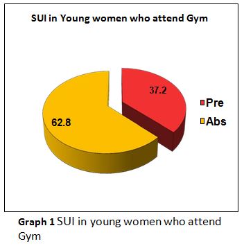
Result: This study enrolled 500 young women with an average age of 22.77. 37.2% of the young women attending the gym had SUI while doing high-impact workouts.

Conclusion: The study reported that there is a risk of stress urinary incontinence during high impact exercises in young women. Both married and unmarried young women experienced leakage during high impact exercises. The finding suggests that continuous high-impact exercise may result in chronic mechanical stress of the pelvic floor.

Keywords: High impact exercises; Stress urinary incontinence; ICIQ-SF score; QUID score.

Received on 27th July 2021, Revised on 12th August 2021, Accepted on 28th August  2021, DOI:10.36678/IJMAES.2021.V07I03.006

INTRODUCTION

Urinary incontinence (UI) is defined by the World Health Organization (WHO) and the International Continence Society (ICS) as an unintentional flow of urine via the urethra, and it is regarded as a concern for one’s health, social well-being, and hygiene. Urinary incontinence can be classified into three categories, according to the standardization steering committee (SSC). Stress urinary incontinence(SUI) or effort urinary incont-inence, urge urinary incontinence (UUI), and mixed urinary incontinence are the three types of incontinence (MUI)1.

The  urgency urinary incontinence (UUI), it is described as the uncontrolled loss of urine that occurs in response to a sense of urgency, there are two other clinical presentations to consider: mixed urinary incontinence (MUI), which is described as a relationship between urgency and leaking during physical exertion and SUI, which is characterized as any symptom of uncontrolled loss of urine that occurs following physical efforts, such as sneezing and coughing, or during any other stressful situation2.

Among these types of incontinence, SUIis the most common, with prevalence rates ranging from 10% to 55% in women aged 15 to 64 years.3The significant risk factors can be classified as predisposing factors, such as family history, supporting factors, such as sports participation, and aggravating factors, such as obesity and natural delivery with neuromuscular impairment. In this scenario, women who engage in physical activity, particularly impact sports, have a higher prevalence of urine incontinence. According to a study, 47% of women who regularly exercise may suffer from urine leakage. Numerous individuals link this issue to pregnancy and childbirth. However, 25–28% of non-pregnant high school and collegiate athletes develop stress urine incontinence4.

These percentages are even more remarkable in athletics that dramatically increase intra-pelvic pressure, such as gymnastics and trampoline, where between 60% and 80% of players report incontinence.”The proportion of urinary leakage in the different sports was gymnastic 56%, ballet 43%, aerobics 40%, badminton 31%, volleyball 30% athletics 25%, handball 21% and basketball 17%.”5Jumping was the most probable action to result in leakage. The majority of the time, SUI is the result of a malfunctioning pelvic floor. Urine leakage is a widespread issue, even among young females, and irrespective of age, between 15% and 30% of women have negative consequences from urinary incont-inence in all sectors of life, and including a decline in life quality. SUI affects one in every three women at some point in their lives and is more prevalent in parous women than in nulliparous women6,7.

Urinary incontinence is resulted by a complicated synchronization of the bladder, urethra, pelvic floor muscles, and ligaments, which occurs most frequently during activity when abdominal pressure increases.The magnitude and duration of high-impact, frequent exercise may contribute to pelvic floor muscle fatigue and, as a result, incontinence. Raises in intra-abdominal pressure are evenly transferred to the bladder, bladder base, and urethra in an optimally supported urogenital tract. Increased downward direct pressure caused by coughing, laughing, sneezing, and Valsalva manoeuvreis balanced incontinent women by ancillary tissue tone produced by the levator ani muscle and vaginal connective tissue. However, when descending forces are not resisted in those with a compromised supporting “backboard, “funnelling of the ureterovesical junction occurs, resulting in an open urethra and subsequent urine leakage 8,9,10.

High impact activities are those that need both feet to leave the ground simultaneously, such as sprinting, jumping jacks, plyometrics, some stepping aerobics, and some cardio dancing that requires hopping. These activities should be kept for those who already have a basic level of fitness and are at minimal risk of developing joint problems, as they carry a significant risk of damage, particularly to the ankle, knee, and hip joints, and also the spine.Fit and active women are more prone to experience SUI. Numerous researches have demonstrated that activities requiringmuch physical exertion andhigh impact exercise can significantly increaseintra-abdominal pressure. It may overburden the pelvic organs, resulting in injury to the muscles that brace these organs. Within this setting, exercise becomes a contributor to the progression of urinary incontinence in women, particularly those without a history of childbirth or pregnancy11,12.

Regular exercise was already recommended to people of any age for its health benefits and as a means of compensating for an ina-ctive lifestyle that can develop obesity, muscle weakness, and postural difficulties. Men and women work out at gyms with this goal in mind, oblivious to the fact that when performing the activities, the peripheral and interior muscles are involved and may be harmed if the activities are performed inappropriately. At some stage of life, all women will have stress urinary incontinence. Unfortunately, many of them “live with” the problem, either because they are too ashamed to ask for help or because they believe it is a “natural” aspect of ageing and having children13,14.

There has been little research on the influence of high-impact workout on the female lower urinary system. The more effective treatment for SUI is surgery. In women and individuals with minor symptoms, conservative treatment is now suggested as the first line of defence. Conventional non-surgical treatmentsinclude lifestyle changes, bladder strengthening and conditioning, pelvic floor muscle workout, biofeedback, and activating pelvic floor by applying a small voltage.

Kegel workouts are the most common approach toreinforcing the pelvic floor muscles because they are non-invasive and do not require vaginal weights or cones. Arnold Kegel, an American gynaecologist, characterised them for the first time in 1948. They seem to be the most cost-efficient treatment option and are distinguished from other therapies because they may be performed independently by the patient at any time and location while performing other tasks without requiring regular hospital trips. Simply instructing the patient on how to tense their pelvic floor muscles is all that is required. Kegel exercise consistently strengthens the pelvic floor, according to the majority of studies15.

 By conducting the survey,it tries to find out the prevalence of SUI in young women who participate in high impact exercises at the gym in Kannur and Kozhikode districts in Kerala and also give awareness to the public regarding the significance of pelvic floor strengthening exercises and the relevance of the same in having healthy adultlife.

MATERIALS AND METHODOLOGY

Five hundred young women were selected by the proper screening and fulfilling the inclusive criteria: a) Young women aged 20-25 years, b) Young women who satisfied specific questionnaire criteria about the type of activity in the gym, c) Healthy and sexually active women, d) Willingness to participate, e) Young women who attend gym and exclusion criteria f) Handicapped young women, g) Surgical treatment for gynaecological and urological illness, h) Infection of the urinary tract, i) Diabetics Mellitus, j) Respiratory disease, k) Incomplete questionnaire, l) Refusal to take part in the research, m) BMI above 30, n) Combination of multiplesports.

Before the study, the purpose and procedure were explained to the participants, and their consent wasobtained.  The participant’s height and weight were measured to calculate BMI. The 500 participants who performed high impact exercises were given two questionnaires.The “ICIQ-SF (International Consultation on Incontinence Questionnaire-Short Form)” questionnaire contains four primary items in which respondents are asked to rate their symptoms during the previous four weeks. The first two items are demographic, while the final one is self-diagnostic. The QUID (the questionnaire for urinary incontinence diagnosis) has six questions regarding urinary incontinence, in which three items consist of SUI and the remaining three items consist of urge incontinence symptoms. Both questionnaires were given to each participantand recorded.

RESULT

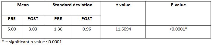
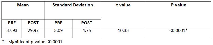
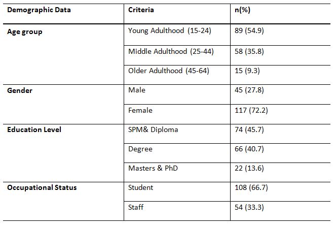
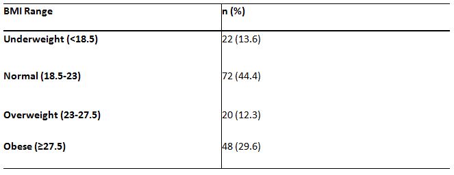
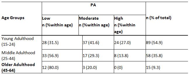
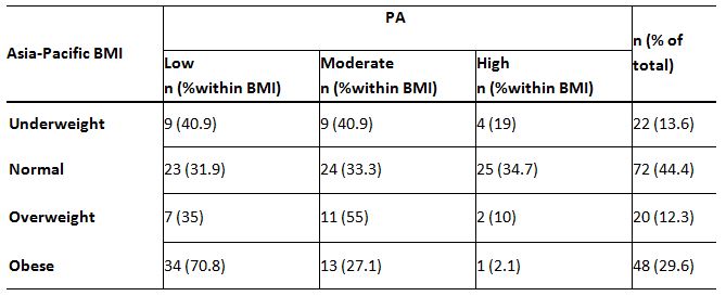
This study enrolled 500 young women with an average age of 22.77.37.2% of the young women attending the gym had stress urinary incontinence while doing high-impact exercises. The majority of the young women having SUI were in the overweight category. The p-value is less than 0.001. In the QUID score, the relation between age and SUI is positive. The SUI is seen to increase with age. However, since the p-value is greater than 0.05, the coefficient is not statistically significant, and the relation between BMI and SUIis positive.

The SUI is seen increasing with BMI. Since the p-value is less than 0.001, the coefficient is statistically significant. According to the ICIQ SF score, the relationship between age and SUI is positive, and the SUI also increases with age. However, since the p-value is greater than 0.05, the coefficient is not statistically significant, and the relation between BMI and SUIis positive. The SUIis seen increasing with BMI. However, the p-value is greater than 0.05; the coefficient is not statistically significant.

DISCUSSION

The research aimed to determine SUI prevalence in young women who go to the gym and participate in high-impact workouts. “According to the World Health Organization (WHO) and the International Continence Society (ICS), urinary incontinence is defined as the involuntary flow of urine via the urethra and is a cause for concern on health, social, and hygiene reasons”.SUI, UUI, and MUI are the three primary kinds of urine incontinence. In a nutshell, SUI is defined as the loss of urine due tohigh intra-abdominal pressure and rise in intravesical pressure beyond the urethral closure limit. This scenario can arise as a result of coughing, sneezing, or jumping. Urinary incontinence is more frequently diagnosed in women than males, and it was anticipated to impact almost 420 million individuals globally in 2018. According to recent data, women have urinary incontinence twice as frequently as males. This condition affects approximately 20-30% of young women, 30-40% of women in middle age, and up to 50% of old aged women.

High impact activities are those that require both feet to leave the ground simultaneously, such as sprinting, jumping jacks, plyometrics, some step aerobics, and some cardio dancing that incorporates leaping. These types of exercise should be confined to individuals who already have a baseline level of fitness and are not at risk of developing joint problems, as they increase the risk of damage, particularly to the ankle, knee, and hip joints, as well as the spine11. Numerous women incorporate recreational physical activity into their daily routines in order to ensure a healthy lifestyle. Physical activity has long been known to benefit blood pressure, weight loss, diabetes, and hyper cholesterolemia. Men and women work out at gyms with this goal in mind, paying little attention to how they are doing so. The impact of physical activity on normal physiology of bladder, on the other hand, is not well recognized.

According to Celina Fozzati c, this occurs on physical workout when the intra-abdominal pressure varies. Pelvic floor muscle activity can be harmed by injury, and their malfunction is one of the determinants of the evolution of SUI in adult females.16High impact activities have been known to rupture the endopelvic fascia or the arch tendinous insertion of the pelvic floor muscles. Another possibility is that alterations in the spine’s physiological curvature result in postural changes and modification of the pelvic cavity’s anatomy due to stretch or compression injury to the pelvic floor muscle.

In this study, 500 young women are selected from the various female fitness centres in Kannur and Kozhikode districts in Kerala based on inclusion and exclusion criteria. A validated “ICIQ-SF questionnaire (International Consult-ation on Incontinence Questionnaire-Short Form)” and “QUID (the Questionnaire for Urinary Incontinence Diagnosis)” questionnaire was used for this study. ICIQ SF questionnaire,has four major sections that ask for a rating of symptoms during the previous four weeks. Items 1 and 2 are demographic, and the final item is self-diagnostic.

The QUID is the questionnaire in which six questions regarding urinary incontinence in which three items consist of SUIand the remaining three consist of urge incontinence symptoms. All the subjects were given and explained the questionnaire. From the questionnaire outcome, we obtained the incidence of SUIin young women who perform high impact exercises atthe gym.

SUI develops when a weakened urethral sphincter cannot withstand the discharge of urine from the bladder during instances of increasing intra-abdominal pressure. “The respective elements affect to urethral continence maintenance: passive urethral closure and coaptation, a critical urethral length, maintenance of the bladder muscle and proximal urethra in their normal anatomic positions, and adaptive changes to the urethra during periods of increased intra-abdominal pressure17.

According to Kari BO, high-impact physical activity increases intra-abdominal pressure. Suppose the muscles of lower pelvic region are unable to co-contract rapidly andsufficiently powerfully to resist this immense pressure or endure the ground compressive force, the levator hiatus may widen, extending and weakening the muscles and increasing the risk of urinary incontinence18.

Epidemiological research on urine incontinence indicates that the disorder is two to three times more prevalent in women. Thus, urinary incontinence can be considered a specific aspect of ageing when it is not reported in women of all ages, cultures, and races, contrary to popular belief that it is more prevalent in the old population, creating a global problem.

According to Hannestad, the frequency grows steadily with age, reaching a significant high in middle age and continuing to rise continuously after age 65. The kind of incontinence may vary with age; some research indicates that SUI is more prevalent in women under the age of 60. According to the survey, the majority of cases were documented among young women under the age of 25. High BMI is a substantial and unique risk factor for SUI, according to Navneet margon10.Evidence suggests that both desire and SUI prevalence increase proportionately with BMI. Thus, a rise in intraabdominal pressure associated with higher BMI results in a correspondingly increased intravesical pressure, which overcomes urethral closure pressure and results in incontinence.

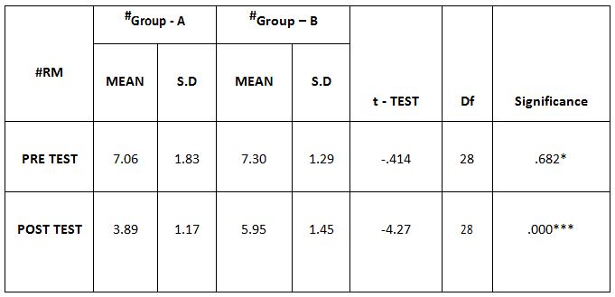
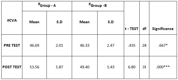
In this study, 37.2% of the young women attending the gym had stress urinary incontinence. According to QUID and ICIQ – SF scoring, the relation between age and stress incontinence is positive. The SUI is seen to increase with age. Nevertheless, since the p-valueis greater than 0.05, the coefficient is not statistically significant. The relation between BMI and SUI is positive.

According to ICIQ –SF score SUI seen in increasing with BMI, the p-value is greater than 0.05; the coefficient is not statistically significant. But in the QUID score, SUI increase with BMI. Since the p-value is less than 0.001, the coefficient is statistically significant.

Ethical Approval: Ethical clearance has been obtained from the Ethical Committee of Cooperative Institute of Health Sciences, with reference number: 01/2018/MPT Musculo-skeletal & Sports/CIHS, dated: 12/04/2019, Kozhikode, Kerala.

Conflicts of Interest: No conflict of interest to conduct and publish this article was reported throughout the study.

Fund for the Study: This study is self-funded.

Limitations: The study was conducted in a small sample size. All the data were collected subjectively, which may introduce an error that treats the study’s reliability. No pilot study was conducted.

CONCLUSION

 The study concluded that SUI is a potential danger in young women who engage in high-impact workouts. Both married and unmarried young women reported experiencing leakage throughout high-impact activities.

The study’s findings will assist young women to engage in high-impact activities to strengthen their pelvic floor muscles and prevent stress urine incontinence.

REFERENCES

  1. Magdelena Weber-Rajek, Agnieszka strac-zynska. Assessment of the effectiveness of pelvic floor muscle training (PFMT) and extracorporeal magnetic innervation (ExMI) in treatment of stress urinary incontinence in women: A Randomized Controlled Trial. Biomed Res Int. 2020 Jan 16; 2020; 1019872.
  2. SoralaTonon Da Luz. Urinary incontinence in physically active young women: preva-lence and related risk factors. Int J Spors Med2017; 38; 937-941.
  3. AratiMahishale, Rafat Khalid Hussain Jamadar. Research Article screening of urinary incontinence in female dancers- A cross sectional study. Research Journal of Obstetrics and Gynecology ISSN 1994-7925 DOI: 10.3923/rjog. 2018;31;35.
  4. Ellen Casey MD. stress urinary incontinence in the female athletes. July 9; 2015.
  5. H. H.Thyssen, L. Clevin,S. Olesen. Urinary incontinence in elite female athletes and dancers. Int Urogynecol J. 2002; 13; 15-17
  6. Magdalena Hagovska, Jan Svihra. Prevalence of urinary incontinence in female performing high impact exercises. Int J Sports Med. 2017; 38; 210-216.
  7. Aletha Silva Caetano, Maria de Consolacao Gomes Cunha. Urinary incontinence and physical activity practice. Esporte Niteroi July/Aug2007; Vol. 13; No 4; pp 245e-248e.
  8. Jodie G. Dakic, Jill Cook, Jean Hay-Smith Pelvic floor disorders stop women exercising: A survey of 4556 symptomatic women. Journal of Science and Medicine in Sport. June 2021; S1440-2440(21)00147-X.
  9. Aletha Silva Caetano, Maria de Consolacao Gomes Cunha. Urinary incontinence and physical activity practice.Esporte,Niteroi July/Aug 2007; Vol.13 no 4.
  10. Navaneet Margon, Bharati karla. stress urinary incontinence what, when, why and then  what. Journal of mid life health, Jul- Dec 2011; Volume 12.
  11. Carls C. The prevalence of stress urinary incontinence in high school and college-age female athletes in the midwest: implications for education and prevention.  Urol Nurs. 2007 Feb; 27 (1); 21-24.
  12. Jyoti S Mandge, ArmaityDehmubed. Study of urinary incontinence affecting quality of life and health seeking behavior in women of an urban slum in Mumbai. Int J community Med Public Health. 2019 Jan; 6(1); 290-292.
  13. Orly Goldstick, Naam Constantini . Urinary incontinace in physically active women and female athletes. BJSM online First, published on May 18, 2013 as 10.1136/ bjsports-2012-091880.
  14. Beta Stach-Lempinen, Clas-HakanNygrad. Is physical activity influenced by urinary incontinence. BJOG: an International Journal of Obstetrics and Gynecology May 2004, Vol.111; pp.475-480.
  15. Seong Hi Park, Chang Bum Kang. Effect of kegel exercise on the management of female stress urinary incontinence. A systemic review of Randomized controlled trials.volume 2014/Article ID640262.
  16. Celina Fozzatti. Prevalence study of stress urinary incontinence in women  who perform high impact exercises. Int Urogynecol J., 2012; 23(12); 1687-1691.
  17. Rovner, E. S., & Wein, A. J. (2004). Treatment options for stress urinary inco-ntinence. Reviews in urology, 6; Suppl 3; S29–S47.
  18. Kari. BO .Is physical activity good or bad for the female pelvic floor?. A narrative review. Sports medicine. (2020). 50; 471-484.
Citation: Kavya Sree.P.P, Saji.V.T(2021). A survey on the effect of high impact exercises on stress urinary incontinence in young women, ijmaes; 7 (3); 1080-1087.

Study on Prevalence of forward head posture among young individuals wearing eye glasses

Manoj Abraham. M1, Meenas Mohamed Sageer2

Author:

1Principal, KG college of Physiotherapy (The Tamil Nadu Dr. M.G.R. Medical University), Thudiyalur Road, Saravanampatti, Coimbatore, Tamilnadu, India,  Email id: manojpt3@gmail.com

Corresponding Author:

2BPT Internee, KG college of Physiotherapy (The Tamil Nadu Dr. M.G.R. Medical University), Thudiyalur Road, Saravanampatti, Coimbatore, Tamilnadu, India,   Email id: skmeenaskm@gmail.com

ABSTRACT

Background: Wearing eyeglasses have a greater influence on adapting to abnormal head and neck posture. As our sensory system is created so efficiently where any obstruction to the visual field will be compensated by body posture. Using an eyeglass provide a  smaller visual field leading to a chronic postural adjustment of the neck causing deep neck flexor weakness and eventually adapting to forward head posture.Aim of the study was to spot the prevalence of forward head posture on people wearing eyeglasses.

Methodology: The study was conducted among 106 eyeglass wearing populations of age groups 19-38. The subjects were assessed subjectively by giving a self-designed questionnaire and objectively the Craniovertebral angle was measured using the photographic method at KG College of physiotherapy outpatient department, Coimbatore.

Result: The study found that 75% of the population was affected by forward head posture and there is a moderate correlation (r value= 0.64) of hours of wearing eyeglasses and craniovertebral angle.

Conclusion:  Supported the result obtained, it is found that there is a high prevalence of forward head posture in people wearing eyeglasses and it depends on the duration of wearing eyeglasses. Thus, the finding of this study revealed that it is important to comprehend that wearing eyeglasses harms head and neck posture and also the importance of assessing visual devices when patient complaints of neck pain and disability.

Keyword: Forward head posture; Eyeglasses; Craniovertebral angle.

Received on 29th July 2021, Revised on 10th August 2021, Accepted on 26th August  2021, DOI:10.36678/IJMAES.2021.V07I03.005

INTRODUCTION

Proper posture is the state of absolute balance with a negligible amount of stress and strain.  Although it is desirable people are unsuccessful in achieving it1.This is probably due to muscle elongation or shortening which leads to inefficiency of movement, disability and pain2.

Forward head posture (FHP) is one of the increasingly prevalent postural deviations. It is the main potential risk factor for various musculoskeletal and neurological problems even when there is no pain or disability at the present. The prolonged placement of the head anterior to the body’s centre of gravity is one of the main etiologies for forward head posture3. Forward head posture is generated due to shortness of the cervical extensors and pectoralis muscles and weakness in the deep cervical flexor muscles and mid-thoracic scapular retractors4. One of the common methods to access head posture is by measuring craniovertebral angle (CVA) 5.

In most of the low and middle-income countries eyeglasses are still widely used, even after the arrival of latest treatment because it is effective, safe and economically feasible1. Lately, it had been found that wearing an eyeglass alters viewing distance and gaze angle which influences the body posture resulting in future risk factors6. An activity that requires eyeglasses like reading will be done efficiently, only by adapting to small compensatory postures. This is done for making the line of vision perfectly aligned to the focusing object7.

Deep neck flexors (DNF) muscles of the neck play a crucial role in maintaining a stable position of head over cervical spine8. Most of the time the biomechanical movement of the joint is interrupted by prolonged exposure to load on craniovertebral extensor muscles and the surrounding non-contractile structure and this increased stress can cause postural changes and eventually edge onto musculoskeletal damage or pain9.

Previously, there were many studies done to find out the risk factor of forward head posture like long time usage of smart phones or long time desktops working with poor posture. However, in India only few studies have been aimed to find out the relation between the usage of eyeglasses and posture. Hence this study is to find out the proportion at which a very simple and unavoidable material employed in day to day life is affecting one’s body posture which leads to greater expenditure of money and time for the rehabilitation purpose.

Objective of the study: The objective of the study is to identify the prevalence of forward head posture on the person wearing eyeglasses and to find the correlation of craniovertebral angle with hours of wearing eyeglasses.

METHODOLOGY

This is a cross-sectional study design conducted by accessing subjects from the KGISL campus for duration of 3 months. The sampling method for this study is random sampling. The sample size was calculated by using an app named epi info statistical calculator and 106 participants fulfilling inclusion criteria were included.

Inclusion criteria: Voluntary participants of the age group of 19-38 years who use any form of eyeglasses for more than 2 years10.

Exclusion criteria: Any history of prevailing neck pain or congenital abnormality of neck and participant who do regular exercise are excluded. Participants who are not regular users of eyeglasses or who have language or cognitive deficits were also excluded3.

Outcome measure: The outcome measure used here is a self-designed questionnaire for collecting demographic data and the photographic method for accessing craniovertebral angle.

Procedure: The participants were asked to assemble at the physiotherapy outpatient department at the KGISL campus and the following procedure was done.All the subjects were informed about the procedure and written consent were obtained before taking photographs for accessing CVA.

A self-designed questionnaire was created, the first part of which focused on demographic details and details about the work, the second part included questions of social factors and the third part included details about wearing eyeglasses.

This Questionnaire was validated by 3 senior physiotherapists who are involved in occupational health research. The questionnaire was distributed to every individual participant, the questionnaire was explained and participants were given two days to complete the same.

Figure 1. Photographic method

This method is used to assess forward head posture by finding the craniovertebral angle which is normally 49.9 degrees. Photographic method is used in this study to find CVA, which has a reliability of > 0.972. It is also cost-effective, gives accurate angle measurement and has fewer errors. 

First, adhesive markers were placed on the tragus of the ear, and the 7th cervical spinous process.  A mobile (iPhone of 12 megapixels) was placed at a distance of 100 cm on a tripod stand. Participants were made to stand in such a way their side faces the camera and were asked to focus on an object in front of them. The subjects were asked to do the neck movements a few times before achieving the standing neck resting position and focusing on the target object. The necessity of maintaining a natural position before the photography was explained to the participants. Then, three sagittal plane photos were taken by the camera.  Repeated photographs aimed at reducing bias due to participant’s tension during photography capturing as well as to overcome the difference between measurements because of postural swaying.

The photos are then transferred to an application; angle measure and then the C7 spinous process was marked as the vertex of the angle. Two lines were drawn one joining the C7 spinous process and tragus of the ear, another line passing horizontally through the C7 spinous process. The angle formed by these two lines was marked as CVA.

Appropriate information collected was entered in the data collecting sheet for purpose of statistical analysis. Once the procedure was over a ‘thank you’ note along with postural advice and an awareness pamphlet was given to everyone.

Statistical analysis: MS-excel were used to enter the data and find the result. Descriptive statistics, Pearson coefficient correlation and prevalence calculation was done and result obtained.

RESULT

By careful examination of 106 participants, descriptive statistics of participants were analyzed and expressed in the table given below.

Prevalence calculation of forward head posture is done, which shows that there is a greater prevalence of forward head posture in participants wearing eyeglasses such that 70 people i.e, 75% of participants were affected with forward head posture.

Correlation of hours of wearing eyeglasses with craniovertebral angle was calculated, the result reveals that there is a moderate positive correlation between them with r value is 0.64 which indicate that the longer the duration of eyeglass usage worst the FHP is. This finding reveals that a widely used eye correction device will eventually become the cause of sustained pain in the future.

DISCUSSION

The purpose of this study is to find the prevalence of forward head posture in people wearing eyeglasses. In this study 106 regular eyeglass users were selected, they were accessed to find the development of forward head posture.

Risk factor identification is an important factor while accessing a person with neck pain or any other postural deviation11. Forward head posture has been linked with neck pain and dysfunction, cervicogenic headache, carpal tunnel syndrome and even an increased falling risk in the elderly12.

Vision problem is a global health concern, especially in children and adolescents 13. As depicted by the Vision Council of America, some sort of vision correction is used by 75% of adults. In which 64% prefer eyeglasses, whereas only 11% prefer contact lenses, with or without frequent use of eyeglasses. Sustained activation of muscle causes calcium ion accumulation which leads to impaired blood flow (Cinderella hypothesis) and again there will be reperfusion of these muscles finally adapting permanent faulty posture. Using eyewear for a prolonged duration can

lead to muscle damage due to sustained low-intensity muscle activity for a prolonged time. Studies have shown that postural adaptation has been caused while viewing a visual target as a result of interaction between the visual and musculoskeletal systems 14.

Larry Sachs et al, found that on an average population there is a significant increase in the degree of forward head posture in the multifocal lens users than non-multifocal lens users and this gives important information for the physiotherapist who treats patient complaining pain over the neck-shoulder region. He also stated in his study that if a patient is having forward head posture and who is a multifocal lens user, then the lens usage is a greater contributing factor to the change in head posture14

A recent study conducted in India claims that DNF endurance was less in subjects wearing bifocal eyeglasses followed by unifocal and no eye glasses, which leads to a greater risk of getting forward head posture especially in people wearing a bifocal lens3.

These changes in posture may lead to a greater risk of musculoskeletal disorder and headache15. Early identification of risk factors leading to permanent postural changes is helpful to take necessary preventive measures.

Even though an eyeglass has risk factors it is unavoidable. Thus, exercise is the only remedy for potential postural deviations. Regular neck exercise is advisable for a person using eyeglass and more studies must be done for the same. Regular postural assessment must be indicated for a regular eyeglass user and rehabilitation to prevent future postural deviation must be started as earlier as possible.

Ethical Concern: This study had no risk factor involved; the study was approved by Institutional ethical committee K G College of physiotherapy, Coimbatore.

Conflicts on Study: The author declares there is no competing interest in publishing this article.

Fund of Study: This is a self-funded study.

Acknowledgement: I thank all the participants for spending their valuable time to answering the questionnaire given and for letting me take the photographs without hesitation.

CONCLUSION

Based on the result obtained, it is found that there is a high prevalence of forward head posture in people wearing eyeglasses. Thus, the finding of this study revealed that it is important to realize wearing eyeglasses has an adverse effect on head and neck posture. This implicates the need for postural correction and regular head and neck exercise in daily life, especially for those people who wear eyeglasses every day.

While assessing any musculoskeletal problems in the neck, checking the details of the optic devices will be helpful to find out the etiologies and assists to derive a proper diagnosis. This small check could be a big move towards reducing the future financial and psychological burden on patient. It can also be a very less invasive public health solution for postural changes on the neck.

This study has put forward the need for further studies searching optometric parameters causing neck disability and to research on the treatment for the same. Further, this study can be done using radiological examination and finding the effect of using each eyeglass like unifocal, multifocal and progressive types on the neck along with the treatment protocol.

REFERENCES

  1. llesh Patel, Sheila K West (2007). Presbyopia: prevalence, impact, and interventions, Community Eye Health.  20(63): 40-41.
  2. Melody tabatabaei, Behrouz Barjasteh Mohebbi, Alireza Rahimi(2017); The impact of 8 weeks selected corrective exercises on neck pain, range of motion in the shoulder and neck of lifesaver women who suffering from forward head posture and myofascial pain syndrome, journal of biomedical research and therapy.  4(6): 1420-1431.
  3. Alka Pawalia, Shabnam Joshi, Vikram Singh Yadav(2019); Can your eye glasses lead to future neck pain and disability, international journal of community medicine and public health. 6(6)- 2402
  4. Susan Armijo-Olivo Jaime Bravo, David J Magee, et al.,(2006) The association between head and cervical posture and temporo-mandibular disorders: A systematic review, Journal of orofacial pain 20(1):9-23.
  5. Ghi-Hwei Kao, T. K. Philip Hwang (2018) Head and Neck Supporting for Seating International Conference on Human-Computer Interaction HCI ; HCI International 2018; Posters’ Extended Abstracts; pp 375-380.
  6. Karen Grimmer (1998) The association between cervical excursion angles and cervical short flexor muscle endurance; Australian Journal of Physiotherapy 1998; Volume 44; Issue 3; Pages 201-207.
  7. Edmondston S J, Sharp M, nawaf Alhabib N, et al.,(2011) Changes in mechanical load and extensor muscle activity in the cervico-thoracic spine induced by sitting posture modification; Ergonomics. 54(2);179-86.
  8. Haejung Lee, Leslei L Nicholson (2005) Neck muscle endurance, self report, and range of motion data from subjects with treated and untreated neck pain. Joint Manipulative Physiological Therapy; 28(1):25-32.
  9. Bae YH, Lee GC (2011) Effect of Motor Control Training with Strengthening Exercises on Pain and Muscle Strength of Patients with Shoulder Impingement Syndrome. J Korean Soc Physiotherapy; 23(6):1–73
  10. Andreas Hartwig et al.,(2007) analysis of head position used by myopes and emmetropes while performing a near-vision reading task. Vision research. 51(14)1712-1717. 
  11. Aliaa Rehan Youssef (2016) photogram-metric quantification of forward head posture is side dependent in healthy participants and patients with mechanical neck pain, international journal of physiotherapy; Vol 3; Issue 3. Pages 326-331.
  12. Jaap H.van Dieën (2006) Pathophysiology of upper extremity muscle disorders; Journal of Electromyography and Kinesiology Volume 16, Issue 1, Pages 1-16.
  13. J smith, J Lewis D Pricard (2005) Physiotherapy exercise programs: Are instructional exercise sheets effective; physiotherapy theory and practice; 21(2) 93-102.
  14. Lary sachs et al.,(1996) the interaction of wearing multifocal lens with head posture and pain; journal of orthopedics and sports physiotherapy; 23(3) 194-9.
  15. Cureton Jr TK (2013) Bodily Posture as an Indicator of Fitness. Res Quarterly Am Assoc Heal Phys Educ Recreat; 348-357.
Citation: Manoj Abraham. M1,  Meenas Mohamed Sageer (2021). Study on prevalence of forward head posture among young individuals wearing eye glasses, ijmaes; 7 (3); 1072-1079

Prevalence of Upper Crossed Syndrome Among Software Professionals

Manoj Abraham. M1, Soumya Murali2

Author:

1Principal, KG college of Physiotherapy (The Tamil Nadu Dr. M.G.R. Medical University), Thudiyalur Road, Saravanampatti, Coimbatore, Tamilnadu, India,  Email id: manojpt3@gmail.com

Corresponding Author:

2BPT Internee, KG college of Physiotherapy (The Tamil Nadu Dr. M.G.R. Medical University), Thudiyalur Road, Saravanampatti, Coimbatore, Tamilnadu, India, Email id: soumyamurali2818@gmail.com

ABSTRACT

Background: Upper crossed syndrome occurs due to muscular imbalance created in opposite muscle groups developing due to postural disturbances. It is presented with the simultaneous tightening of postural muscles and weakening of non-postural muscles in the upper body resulting in limited mobility. The purpose of the study is to determine the prevalence of upper crossed syndrome among software professionals.

Methods: A randomized sampling of 106 software professionals was conducted based on inclusion and exclusion criteria. The research was a cross-sectional study where software professionals of age group 20-40 years having a daily working schedule of minimum of 3 hours and a maximum of 18 hours on computers were included. A self-designed questionnaire and Neck Disability Index were circulated among participants and were assessed by performing special tests.

Results: The prevalence of upper crossed syndrome among the software professionals is 55.6%. There is a positive linear relationship between the development of the upper crossed syndrome and the functional disability among the people and in the Neck Disability Index and the hours of working in front of computer.

Conclusion: This study illustrates that there is a significant prevalence of upper crossed syndrome among software professionals. Neck pain and functional limitation were presented as the main symptom that is in the verge of occurrence.

Keywords: Upper crossed syndrome, musculoskeletal imbalance, neck disability index.

Received on 29th July 2021, Revised on 10th August 2021, Accepted on 26th August  2021, DOI:10.36678/IJMAES.2021.V07I03.004

INTRODUCTION

India has been the vanguard in the cyber world with the development of information techno-logy. Approximately 6 computers per 1000 population and about 18 million personal computers are prevalent in our country. This number is increasing day by day. The booming of the information technology industry in India has led to an increase in the use of computer devices as well as marked the start of a new genre of occupational health problems among software professionals1. It has led to various musculoskeletal discomforts2. The mushroom-ming of the Visual Display Units (VDU) or the Visual Display Terminals (VDT) in the modern office settings has led to the various occupational health problems and musculo-skeletal ailments associated with it3. With the  association of improper posture, excessive muscular tension, and limited mobility results4. The excess time on work demands results in a significant increase in pain, functional limita-tion and fatigue5.

The physiological response from the work has been termed as “workstyle”. Workstyle has been recognized as a coupling factor for the muscular imbalances creating pain and functional limitation dueto the ergonomic factors inthe computer users6. Among the musculoskeletal complaints, the neck was the common site to be affected because of the static posture during work hours in front of the computer7.

While working in a sitting posture for a prolo- nged time, they tend to adopt a forward head posture shifting the head forward to the spinal central line. This is because they find it difficult to maintain the normal posture of the spine during the prolonged work hours of sitting8. For every one inch of forward head posture, there is an increase of additional 10 pounds of weight of head on the spine. Where the poor sitting may result in forward head posture and rounded shoulders due to altered body kinematics9. This forward head posture results in mechanical stress on the neck and cause smuscular imbalances10 where onegroup of muscles gets tightened and the other group gets weakened and inhibited11.

This leads to the weakening of middle, lower trapezius, rhomboid scrossing ventrally with the weakness of significant cervical flexors. Simultaneously, the tightening and over active upper trapezius, levator scapulae crossing dorsally with the tightness of pectoralis major and minor also occur12. This may also cause rounded shoulder, and abnormal postures of shoulder blades. Janda defined this phenom- enon as Upper Crossed Syndrome (UCS)13. Dr. Vladimir Janda coinedthis syndromeas Upper Crossed because when the weakened and tightened muscles form a cross when they are connected in the upper body14.

These muscular imbalances created in upper crossed syndrome in opposite muscle groups develop postural disturbances. This would res- ult in a reduction of the quality of the glenohu- meral joint. This is because the glenoid cavity will become more vertical because of the serratus anterior weakness creating the scapulae for winging and turning15. Sitting with the posture of forward head position creates upper cervical region extension and lower cervical region flexion which will in turn, reduce the muscle fiber length. This will eventually develop extensor torque around the upper cervical joints which will later lead to musculoskeletal imbalances and abnormalities like reduced scapular upward rotation, incre- ased internal rotation, anterior tilt which will eventually make them more difficult to maintain their erect posture of sitting16. This winging, elevated, and abducted scapula creates scapular dyskinesia there by which creates inrounding of shoulders.

Nearly three-fourths of the software profess- ionals were identified with computer- related health problems and musculoskeletal ailments. This is almost a significant proportion of the population of software professionals. This den- otes a need to emphasize a field of concern among the software professionals, their well being, and occupational health. Hence regular health check-ups and ergonomic advice being sensitized regarding the importance of their wellbeing and proper working conditions17.

METHODOLOGY

This was a cross-sectional study that was conducted in the Physiotherapy Outpatient Department, KGiSL Campus, Saravanampatti, Coimbatore. This study included 106 particip- ants who were selected by a simple random- ized sampling method. The study duration was three months. Epistat info was used for sample size calculation.

The objectives of the study were; to identify the occurrence of upper crossed syndrome among software professionals; to correlate the upper crossed syndrome with the functional disability of the individuals and also to correlate the upper crossed syndrome with the working hours infront of computer among software professionals.

Procedure: A written consent is obtained from every individual who signed up for the study. Before the study, a brief instruction was given regarding the research. Individuals selected for the study were instructed and asked to assemble in the K.G Outpatient Department in Coimbatore. Aself-designed questionnaire was distributed, the first section of which question- naire was focused on demographic details (name, age, gender, years of working) and the second section included social factors (personal and past history of any illness or treatment), the third section included the occ-urrence of neck pain(onset, duration and past treatment ), the fourth section consisted of other factors (history of working details and confirmatory tests) after the filling up of ques-tionnaire the subjects were also given the Neck Disability Index (NDI).

Neck disability index (NDI): It is a self-reported condition-specific questionnaire that includes 10 items- pain intensity, personal care, lifting, reading, headache, concentration, work, driving, sleeping, recreation). This questio- nnaire emphasizes how neck pain affects the ability to manage the activities of daily life (ADL).

The questionnaire was validated by 3 senior physiotherapists who are involved in occupational health research. After completion of filling the data, Trapezius and pectoralis tests are performed. The collected data were noted and taken for analysis. After the compl- etion of the procedure, thank you note accom- panied with ergonomic advice and a postural awareness pamphlet was given to subjects.

Inclusion criteria: Age between 20-40 years18, Software professionals with a computer-related work experience of minimum of 6 months18, with duration of working hours taken into consideration whichis fixed as at least 3 hours a day or 15 hours per week1.

Exclusion criteria: previous history of any surgery/underlying pathology/inflammatory di- seases to cervical spine, known history of fractures of cervical spine or tumors18,19.

RESULTS

This study showed that a significant proportion of software professionals in the present study reported that they experience muscular disco-mforts. This may be due to various factors such as the impact of the prolonged working hours in poor posture, involvement of multiple joints in computing tasks, adopting biomechanical and anatomical alignment of body motions of high physiologic cost leading to muscular imbalances gradually.

Table 1. Demographic data and NDIscore

Most of the subjects spent a prolonged time in the office as their working hours showed that 31% of the population worked in front of the computer for 4-8hours and 69% of them had more than 8 hours of working on the computer with a static posture. Due to this static posture, they develop symptoms of neck pain. Out of 54% males and 46% females selected rando- mly, 94% of the population reported neck pain and 6% had no neck pain. 30(28%) of them experienced pain for less than one year, 40 (38%) mentioned that they are experiencing pain for one to two years and 36(34%) population had pain for more than 2 years. It was also discovered that only 82% of the population had proper back-supported well-cushioned chairs and the remaining 18% had no properly back supported chair in the office which can be a potential ergonomic risk for the yielding of symptoms as they work for prolonged hours.

Neck Disability Index values revealed that 11% of them had 0-8% of scoring stating no disability, 37% of them had mild disability scoring 10-28%, 52% of them scored 30-48% had moderate disability. None of the popula- tion had severe or complete disability. Further from the study, it was found that the symptoms and muscular discomforts were most debilitating causing a reduction in Activ- ities of Daily Living(ADL).

The mean value of the software professionals who are on the verge of developing upper crossed syndrome is 1.44±0.49 subjects and the mean value of their working hours is 3.68±0.46 hours. The value of ‘’r’’ value correlating them is 0.60. This is a moderate uphill (positive) linear relationship between the development of the upper crossed syndrome and the hours of working among software professionals in front of the computer.

The mean value of the software professionals who are on the verge of developing upper crossed syndrome is 1.44±0.49 subjects and the mean value of their functional disability in Neck Disability Index scale is 27.35±12.8.The value of “r” correlating them is 0.54. This is a moderate uphill (positive) linear relationship between the development of the upper crossed syndrome and the functional disability among the people on the neck disability index.

DISCUSSION

The study has focused on the prevalence of upper crossed syndrome among software professionals. Christensen Ketal, in 2015 evide- ntly proposed upper crosss yndrome appears to be a simple muscular imbalance but it may impart huge stress on the economy of the country via resulting in functional disability due to neckpain20.

 As derived from this study, work organization plays an important role, especially when ergonomic measures are largely implemented. A study also has reported that the individuals developing upper cross syndrome were some how associated with bad postureor indulge in any activity that makes them adopt a posture of high physiologic cost thereby leading to a muscular imbalance that will yield upper cross syndrome21. Morrisetal, Stated that the correlation between upper cross syndrome and poor posture is relevant and found the subjects suffering from the upper cross syndrome were mostly because of poor posture or due to any those activity that makes them adopt bad posture developing a muscular imbalance that will yield upper cross syndrome22.

As Kwonetal, in a study in 2015 suggested that If proper steps are not taken at the initial moment when identified with neck pain with functional disability this may become an endemic not only in the working population but also in the people who tend to adopt a poor posture and least bothered regarding their posture, it is suggested that there should be a pace in the blow to generate postural awareness in people of all ages and most predominantly the risk groups who are in the verge of the occurrence of upper crossed syndrome gradually23.

After review and knowing that neck pain is the main symptom initiated, Neck Disability Index was used as the outcome measure used to assess the impact of neck pain on activities of daily living (ADLs). Most of them had neck disability associated with muscular imbalances. On studying musculoskeletal symptoms of the upper extremities and the neck with symptom-predicting factors at visual display terminal (VDT) workstations, preventive measures should be focused on neck and shoulder disorders. So, for preventive aspect this study also states that ergonomic  interventions sho-uld  be implemented.

CONCLUSION

It is concluded that most of the software professionals were having muscular imbalances yielding upper crossed syndrome and experie- nced the muscular ailment symptoms of neck pain leading to functional disability. The prevalence of neck pain to be 94.3% and the verge of developing upper crossed syndrome to be found as 55.6% among the software professionals. They were exposed to the risk of adopting poor postures due to extended periods of working hours in a   static posture. This study provides knowledge that ergonomic advice to be educated and postural awareness should be introduced and proper biomechanics to be utilized during work hours to enable the work efficiently and comfortably thus improving productivity.

Limitations And Recommendation: This study does not focus on any treatment to the participants as the present study only consisted of ergonomic advice and was done within a limited geographical location there by decrea-sing generalizability. No diagnostic tests other than trapezius and pectoralis tests are included. Future studies could evaluate the association between the occurrence of Work-Related Musculoskeletal disorders and various psychosocial factors such as high stress, low control among the Software professionals. Radiographic diagnostic procedures can be done for the findings. A similar study can be conducted on a wider age group to find out theage impact on the development of the upper crossed  syndrome. Treatment protocols can be given for the selected population as the present study consisted of ergonomic advice only.

Ethical Approval: Ethical clearance has been obtained from the K.G College of Physiotherapy Ethical Committee.

Conflicts Of Interest: No conflict of interest to conduct and publish this article was reported throughout the study.

Fund For The Study: This study is self-funded.

Acknowledgement: The authors would like to express sincere gratitude and special thanks to all the participants who took part in the research.

REFERENCES

  1. Sharma, A. K., Khera, S., &Khandekar, J. (2006). Computer relatedhealthproblems among information technology professionals in Delhi. Indian journal of community medicine, 31(1); 36.
  2. Talwar, R., Kapoor, R., Puri, K., Bansal, K., & Singh, S. (2009). A study of visual and musculoskeletal health disorders among computer professionals in NCR Delhi. Indian journal of community medicine: official publication of Indian Association of Preventive & Social Medicine, 34(4); 326.
  3. Singh, S., & Wadhwa, J. (2006). Impact of computer workstation design on health of the users. Journal of Human Ecology, 20(3); 165-170.
  4. Moore, M. K. (2004). Upper crossed syndrome and its relationship to cervico-genic headache. Journal of manipulative and physiological therapeu- tics, 27(6), 414-420.
  5. Lundberg, U. (2002). Psychophysiology of work: Stress, gender, endocrine response, and work‐related upper extremity disorders. American journal of industrial medicine, 41(5); 383-392.
  6. Feuerstein,M., Nicholas, R. A., Huang, G. D., Haufler, A. J., Pransky, G., & Robertson, M. (2005). Workstyle: development of a measure of response to work in those with upper extremity pain. Journal of occupational rehabilitation, 15(2); 87-104.
  7. Yu, I. T. S., & Wong, T. W. (1996). Musculoskeletal problems among VDU workers in a Hong Kong bank. Occupational medicine, 46(4); 275-280.
  8. Calliet, R. (1991). Mechanism of Pain in the Neck and From the Neck. Philadelphia: Neck and arm pain, 348-67.
  9. Janda, V. (1993). Muscle strength in relation to muscle length, pain and muscle imbalance. International Perspectives in Physical Therapy, 83-83.
  10. Waris, P. (1979). Occupational cervico brachial syndromes: a review. Scandinavian journal of work, environment & health, 3-14.
  11. Gu, S. Y., Hwangbo, G., & Lee, J. H. (2016). Relationship between position sense and reposition errors according to the degree of upper crossed syndrome. Journal of physical therapy science, 28(2); 438-441.
  12. Arora, D., &Veqar, Z. (2010). Correlation between endurance of deep cervical flexors and lower scapular stabilisers in computer users with chronic neck pain. British Journal of Sports Medicine, 44(Suppl 1);  i11-i11.
  13. Janda, V. (1996). Rehabilitation of the spine. Baltimore: Williams & Wilins. 319-328.
  14. Umasankar Mohanty (2015), Manual therapy for upper crossed syndrome, J Nov el Physiotherapies, 5:4 http://dx.doi. org/10.4172/2165-7025.S1.001.
  15. Patterson, K., & Evans, O. M. (2000). Predictors of Neck and Shoulder Pain in Non-Secretarial Computer Users. Volume 26, Issue 3, September 2000; pp 357-365.
  16. Kang, J. H., et al (2012). The effect of the forward head posture on postural balance in long time computer-based worker. Annals of rehabilitation medicine, 36(1); 98.
  17. Blatter, B. M., &Bongers, P. M. (2002). Duration of computer use and mouse use in relation to musculoskeletal disorders of neck or upper limb. International Journal of Industrial Ergonomics, 30(4-5); 295-306.
  18. Kumar, S., Sambyal, S., &Vij, J. (2013). Analysis of disability and pain in Computer Professionals with Neck Pain. IOSR JNHS, 3; 6-9.
  19. Dahiya, J., & Ravindra, S. (2013). Effect of scapular position in computer professionals with neck pain. International Journal of Science and Research, 4(5); 2075-2080.
  20. Christensen, K. (2000). Manual muscle testing and postural imbalance. Dynamic Chiropratic, 18(24). November 15, 2000, Vol. 18, Issue 24.
  21. Mubeen I, Malik S, Akhtar W, Iqbal M, Asif M, Arshad A, Zia S,Khalid (2016). Prevalence of upper cross syndrome among the medical students of university of lahore. International journal of physio-therapy 3, no. 3 (2016); 381-384.
  22. 22. Morris,C.E.,Bonnefin, D., & Darville, C. (2015). The Torsional Upper Crossed Syndrome: A multi-planar update to Janda’s model, with a case series introduction of the mid-pectoral fascial lesion as an associated etiological factor. Journal of bodywork and movement therapies, 19(4); 681-689.
  23. 23. Kwon, J. W., Son, S. M., & Lee, N. K. (2015). Changes in upper-extremity muscle activities due to head position in subjects with a forward head posture and rounded shoulders. Journal of physical therapy science, 27(6); 1739-1742.
Citation: Manoj Abraham. M1, Soumya Murali(2021).Prevalence of upper crossed syndrome among software professionals , ijmaes; 7 (3); 1061-1071

Efficacy of Heavy Bags With 15% of Body Weight in Teenagers on Cervical and Shoulder Posture Alignment

Prachi Jain1, Syeda Khanam P2, Manjunatha H3

Corresponding Author:

1AssistantProfessor, East Point College of Physiotherapy, Bangalore, Karnataka, India Mail id: prachijphysio@gmail.com

Co-Authors:

2,3Professor, East Point College of Physiotherapy, Bangalore, Karnataka, India

ABSTRACT

Background of the study: This examination clarifies about the normal weight an understudy needs to convey to school. The pinnacle development happens during pubescence and the development of the affixed skeletal framework stops around 16 – 18 years for guys. The greater part of the investigations on the impact of burden carriage has been centered around patches and climbers fully intent on working on the strategies of burden carriage.

Methodology: This is an observational investigation with 50 male subjects included. With static and dynamic stacking with 15% body weight and changes in stride design in the two circumstances were caught. Subjects were on their own control where gauge or dumped pose is contrasted and act under two distinctive trial stacking conditions. Subjects were weighed with and without their school packs on the one set off aligned electronic scale.

Result: The mean upsides of Craniovertebral Angle (CV), Craniohorizontal (CH) point and Sagittal Shoulder act expanded in all trial load conditions in examination with dumped state. The mean upsides of Step Length (SL) decreased in unique stacking when contrasted with dumped condition while it was not appropriate if there should arise an occurrence of static stacking. No huge contrasts were found in CH Variance between the dumped condition and conveying school sacks weighting 15% body weight while in static stacking the CH Variance was expanded to practically twofold when contrasted with no heap and dynamic burden condition.

Conclusion: This experimental study supports that dynamic loading of the student with 15% of body weight, leads to significant change in cervical and shoulder posture compared to static loading with 15% of body weight and unloaded condition.

Keywords: Cranio-Horizontal Angle; Cranio-Vertebral angle; Sagittal Shoulder posture; Strength

Received on 12th July 2021, Revised on 18th August 2021, Accepted on 26th August  2021, DOI:10.36678/IJMAES.2021.V07I03.003

INTRODUCTION

The word rucksack was begat in the U.S. in 1910’s. Rucksack is a proper method of conveying load of spine, intently and evenly while keeping up with dependability. The Backpack is one of the few from of manual burden carriage that gives flexibility and is frequently utilized by climbers, hikers and troopers, just as school kids. However, there is developing public worry that over-burden youngsters and juvenile’s knapsacks might prompt the improvement of back torment and other musculoskeletal wounds. Data got from these investigations may duplicate to school kids. Stance in juvenile can be influenced by both inward and outer impacts, which might make young adult more vulnerable to injury. Contemplating postural reactions to burdens will help us to understanding the effect of conveying school packs on kids. At the point when burden is situated back to the body as knapsack it changes pose on account of changes to focus of gravity1, 2

The body attempts to keep the focal point of gravity between feet, so with a bag, the storage compartment is in a more forward position, putting unusual power on the spine, load conveyed in a backpack shift the focal point of gravity behind the body to equalize, the focal point of the gravity of the body in addition to the load is moved back over base of the feet. The specialists demonstrated that conveying backpack lead protrusion of head to the forward direction. These progressions in arrangement of the neck can produces strain of cervical joints 3, 4.

Conveying back loads by youngsters has been connected with spinal agony, and the measure of postural change created by load carriage has been utilized as a proportion of the possibility to cause tissue harm. Back torment in kids gives off an impression of being more normal than was recently suspected. Studies have shown that 10% to 30% of sound youngsters experience back torment by their teen. Consequently examination to postural reactions to stack conveying will assist us with understanding the effect of school knapsack on youngsters5-7.

To decrease the event of musculoskeletal agony a proactive, preventive methodology utilizing ergonomics mediation has been proposed by certain scientists. For instance, school packs plan, school spot, furniture, and apparatuses. Managerial controls are choices made by school staff, medical care experts and others (specialists, school overseers, and guardians or parental figures) to lessen the term, recurrence and seriousness openness to existing risks. They additionally pass on the dangers set up however endeavors to decrease the impacts on the young (for example ensuring the heaviness of the rucksack doesn’t surpass 15% of the body weight, checking the adolescent stance when wearing the knapsack). Work practice controls are self-guided, self-started systems utilized by understudies to guarantee protected and legitimate methods while doing exercises (for example utilizing the two ties, affixing the lashes yet not very firmly). This load of methodologies or controls target decreasing the weight on musculoskeletal framework8, 9.

The limitation of the most extreme load to 15% of the body weight is one of the fundamental controls. Anyway some different creators have suggested that sack weight ought not to surpass 15% of the body weight. In this investigation we are attempting to decide if the heaviness of the rucksack (15% of body weight), its situation on the spine or time conveyed influenced youths cervical and shoulder act. None of the scientists till date have examined reactions of cervical and shoulder act after static stacking and after unique exercises with 15% of body weight. In addition, attributable to anthropometrical contrasts among western and Indian offspring of comparable examination done there are not straightforwardly appropriate to Indian kids 10.

Purpose of the Study: There is need of this investigation so it can give us data about the normal weight a kid needs to convey to school. Subsequently if preventive measures can be acquaint now with the respect with safe burden carriage in undergrads, it won’t just serve to item youngsters while they are as yet growing, yet will likewise guarantee, that the standards they adapt now are brought through to work place as grown-ups and to address the very example deviations which happen in the youngsters because of weighty burden.

Aim & Objectives : Objectives of the study were to examine the changes in Cervical Lumber & Shoulder posture in college children with Dynamic Loading (15% of body wt.); also, to examine the changes in cervical Lumber & Shoulder posture in college children with static Loading (15% body wt.). And to analyses the gait pattern after back loading in college children.

Growth: Growth applies powers to the spine, the extent of which differs with the pace of development. Since the development speed is most elevated in newborn child and youths, it is normal that the subsequent powers applied to the spine are most noteworthy at these ages. Despite the fact that they are not more noteworthy, the powers that outcome from development might change spinal design since they are applied throughout extensive stretches of time. In ordinary spinal development, foremost and back development and side to side development are adjusted. The outcome is stretching of the spinal segment with moderately little change in its gross forms. Assuming, nonetheless, a pathology condition causes deviated development, the power vectors change and cause deformation.

Adaptability The pediatric spine can adjust to applied anxieties substantially more promptly than can the grown-up spine. “This is identified with development potential, the lower modulus of flexibility, and unmistakable rebuilding capacity.

Malleability: as well as being versatile, which suggests in dynamic interaction, the pediatric spine likewise is generously pliable. Pliability, a uninvolved interaction, suggests that the spine might be disfigured with the use of powers outside to the spinal segment.

Hypermobility: The physiologic scope of movement of the pediatric spine is extensively more noteworthy than that of the grown-up spines. This is the consequence of contrasts in ligamentous limitations and direction of the feature joint.

Weak Growth Plate:  The development plate is the most fragile connection in the hub skeleton when it is exposed to tractable powers. This has significant ramifications for the sorts of injury probably going to happen in the pediatric spine. Odontoid wounds typically happen through the actual plate, situated close to the foundation of the odontoid cycle.

METHODOLOGY

Total 50 students aged between 17-20 years of BPT First Year was participated in the study. Participants with fever, systemic illness, cervical injuries, scoliosis or Kyphosis or congenital deformity were excluded from this study. Participants with complaint of pain were stopped  for  testing procedure.

Inclusion Criteria: Students should be aged between 17-20 yrs. Height, Body Weight should almost be similar, weight of the bag should be equal, lifestyle variation.

Exclusion Criteria:  Scoliosis or Kyphosis, Injury, Fever, Systemic illness, any congenital deformity.

Study Procedure: Participants were weighed with and without their school packs on the one set off adjusted electronic scale (Beurer scale, precise to be inside 0.1 kg to 120 kg). Standing stature was estimated against an estimating tape got to divider. One school pack was utilized for every one of the exploratory conditions. The school sack had two movable cushioned shoulder ties, two compartments and no midriff or chest pressure lashes. Scopes of loads of 2 kg, 1 kg, 500 gm, 100gm, and 50 gm were utilized for the exploratory burden conditions.

The element of loads repeated regular instruction material. One Cannon 7.1 Mega pixels advanced camera was utilized to take actually photos of subject’s sagittal stance. Stand remain with a soul meter level was utilized for mounting the camera. Proportions of cervical and shoulder act were determined structure advanced photos utilizing the digitizing programming (Image Tool UTHCSA variant 3.0 University of Texas Health Service Center, San Antonio, TX).

Design of the study: This was an observational study to compare 2 test load conditions  in static and dynamic stage with 15% body weight and  were tried noticing the progressions in step design in the two conditions. Subjects were their own control where benchmark or dumped act is contrasted and act under two distinctive exploratory stacking conditions.

Techniques: Clothing was reworked so that shoulders were uncovered. With the subjects in standing stance cement markers were put on four physical tourist spots.

Spinous process of the seventh cervical vertebrae, Midpoint between greater tuberosity of humerus bone,  back part of acromion of the scapula, Outer canthus of right eye and right tragus, were noted and were approached to stand serenely with their arms close by in ordinary standing stance.

They were approached to put their weight equitably on the two feet. The horizontal malleoli were put between equal lines, which are opposite to the front facing planes. The subjects gazed straight ahead. Camera was put two meters from the subject’s right side. Photo was taken inside 5 second of taking on the position.

The photos of the subjects were taken without school bag; it was 0% body weight, 15% body weight applied for Static loading and for dynamic activities. Information was examined by advanced programming Image Tool UTHCS version 3.0, University of Texas Service Center, San Antonio.

Data Analysis: Examination of postural points after powerful exercises is finished with static loading with 15% body weight and with 0% body weight. The meaning of changes in information was assessed utilizing rehashed measure examination of difference on each point with which arranged differentiation were made of the dumped condition with every one of two other stacked condition. The study was considered as significant with if p<0.05. The Gait was observed at carrying school bag more than two shoulders comparable to 15% body weight with static and dynamic exercises.

Graph 1: The Cranio-horizontal angle increases in the Group A i.e.  Without static loading and it reduces gradually once it is loaded and further more it reduces in the dynamic loading subjects when they start walking.
Graph 2: Cranio vertebral angle increases in static loading comparatively more than unloaded group and dynamic loading.
Graph 3: Shoulder sagittal angle increases in static loading comparatively more than unloaded group and dynamic loading
Graph 4: The step length (SL) changes in group C than group A, which is unloaded static group.

RESULT

The mean stature and mean load of the subjects enlisted were 157.5cms, 54.34 kg separately. The mean load of the bag which kid conveyed to school was observed as 8.151 kg which was 15% of their body weight. Benchmark esteems were got ten by estimating Craniovertebral Angle, Craniohorizontal Angle, Sagittal Shoulder stance and step length on the dumped condition with 0% of their body weight.

The mean upsides of craniovertebral point diminished in a trial loaded condition in examination with dumped condition. The mean worth of CVA in the dumped state was 40.718 = 4.991, though the mean upsides of CVA while static stacking with 15% of body weight and after unique exercises with 15% of body weight were 43.994= 3.866 and 40.542=2.910 individ-ually. Huge contrasts were found by rehashed proportion of difference (ANOVA) in the CVA between dumped state, static stacking and after powerful exercises with p esteem 0.018 which is more than 0.0001.

Pair insightful correlation was between loaded state and static loading with 15% body weight shows that there is contrast with p esteem 0.0001 and furthermore critical distinction between dumped state and after powerful exercises act with p esteem 0.034. There was no critical contrast was found between static stacking states and after unique stacking exercises pose with 15% body weight load for CVA as p esteem is more noteworthy than 0.0001.

The mean upsides of Craniohorizontal point expanded in every one of the two trial load conditions in examination with dumped state. The mean worth of CHA in the dumped state was 16.61 = 2.75, though the mean upsides of CHA while static stacking and after unique exercises with 15% body weight were 23.186 = 3.693 and 20.4 = 2.456 individually. No huge contrasts was found in CHA between the dumped condition and conveying school sacks weighting 15% body weight while static stacking and after powerful exercises act.

The mean worth of sagittal shoulder act increments in every one of the two trial loads conditions in correlation with dumped state. The mean worth of sagittal shoulder act in the dumped state was 38.324 = 6.323, though the mean upsides of sagittal shoulder pose while static stacking and after unique exercises with 15% body weight were 34.68 = 5.977 and 40.75 = 8.850 separately.

The mean worth of step length diminishes in all trial loads conditions in examination with dumped state. The mean worth of step length in the dumped state was 19.06 = 3.436, though the mean upsides of step length while static stacking isn’t pertinent and after powerful exercises with 15% body weight was 16.84 = 3.683.

DISCUSSION

Shruti. R. Iyer in their examination tracked down that Indian youngsters convey school bags gauging 18.5% of their body weight. Pascoe et al7 in their examination done in America tracked down that mean load of school bag conveyed by school kids in the age gathering of 11-13 years was 17% of their body weight11.

Likewise Negrini et al in their examination done in Italy tracked down that normal burden conveyed by school kids matured 11.29=0.33 was 9.3 kg, which was determined to 22% of their body weight. Conversely, the heaviness of school pack communicated in level of body weight in this examination was observed to be heavier than detailed by Forjuoh SN et al in their investigation done in Texas (6.2% among kindergarteners and 12% among fifth graders)12

The consequence of this examination uncovered that the greater part of the Indian kids in the age gathering of 17-20 years conveyed school pack weighing between 15% – 18% of their body weight. The mean worth of weight of the school pack conveyed by youngsters was observed to be 8.151 kg which is observed to be 15% of their body weight. The heaviness of the school pack communicated in level of body weight was observed to be reliable with studies done by Shruti. R. Iyer5 and Pascoe et al13.

Likewise J.K Whitefield et al in their examination done in New Zealand College detailed 13.2% of body weight for 3rd grade and 10.2% for 6th grade. Craniovertebral point gives an assessment of head on upper back. A little point shows more forward head position. It has been tracked down that more modest the CVA point is related with migraines in females51. Additionally change in arrangement of neck can deliver strain of cervical joints and delicate tissues just as imbalanced muscle execution. Head act immediately affects the situation of mandible and can prompt temporo-mandibular joint brokenness, and gulping difficulties14.

Ethical clearance: There was no risk of conducting this study.Ethical clearance was obtained from the ethical committee of Institute of Applied Medicine and Research, under the Chaudhary Charan Singh University, Meerut with approval letter dated 15thApril 2011.

Conflicts of Interest: There is no conflict of interest to conduct this study.

Fund for the study: This is self-funded study.

CONCLUSION

The consequence of the investigation upholds the exploratory theory that powerful stacking the understudy with 15% of body weight prompts huge change in cervical and shoulder pose when contrasted with static stacking with 15% of body weight and dumped condition, Significant change in cervical and shoulder showed by decline in craniovertebral point and expansion in sagittal shoulder act was found in unique stacking and static stacking when contrasted with dumped act.

We have likewise tracked down that 5 minutes of dynamic exercises with 15% of body weight produce huge change in craniovertebral and sagittal shoulder pose when contrasted with dumped act. Consequently, suggesting that school sack gauging 15% of body weight would be excessively weighty for the Indian school youngsters matured 17-20 to have the option to keep up with their ordinary cervical and shoulder pose arrangement.

REFERENCES

1. Knapik J, Harman E, Reynolds K. Load carriage using packs: a review of physiological, biomechanical and medical aspects. Applied ergonomics. 1996 Jun 1;27(3); 207-16.

2.  Voll HJ, Klimt F. Strain in children caused by carrying school bags (author’s transl). Das Offentliche Gesundheitswesen. 1977 Jul 1;39(7); 369-78.

3. Oliveira R, Cabri JM. Low back pain in young people: cross-sectional study in Lisbon. In AIESEP 2005 World Congress-” Active lifestyles: the impact of education and sport”, 2006; pp.233-237.

4.  Taimela S, Kujala UM, Salminen J J, Viljanen T. The prevalence of low back pain among children and adolescents: a nationwide, cohort-based questionnaire survey in Finland. Spine. 1997 May 15;22(10);1132-6.

5.  Iyer SR. An ergonomic study of chronic musculoskeletal pain in collegechildren. The Indian Journal of Pediatrics. 2001 Oct;68(10); 937-41.

6.  Negrini S, Carabalona R. Backpacks on! School children’s perceptions of load, associations with back pain and factors determining the load. Spine. 2002 Jan 15;27(2); 187-95.

7.  Pascoe DD, Pascoe DE, Wang YT, Shim DM, Kim CK. Influence of carrying book bags on gait cycle and posture of youths. Ergonomics. 1997 Jun 1;40(6); 631-40.

8. Whittfield JK, Legg SJ, Hedderley DI. The weight and use of school bags in New Zealand secondary colleges. Ergonomics. 2001 Jul 1; 44(9); 819-24.

9.Chansirinukor W, Wilson D, Grimmer K, Dansie B. Effects of backpacks on students: measurement of cervical and shoulder posture. Australian Journal of physio-therapy. 2001 Jan 1;47(2); 110-6.

10. S.I. Weinsein, Garry M. Banks and Ensor E. Transfeldt. 1994. The Pediarics Spine: Principles and Practice. Raven Press. Ltd. New York.

11. Raine S, Twomey LT. Head and shoulder posture variations in 160 asymptomatic women and men. Archives of physical medicine and rehabilitation. 1997 Nov 1; 78(11);1215-23.

12. Watson DH, Trott PH. Cervical headache: an investigation of natural head posture and upper cervical flexor muscle performance. Cephalalgia. 1993 Aug; 13(4); 272-84.

13. Darling DW, Kraus S, Glasheen-Wray MB. Relationship of head posture and the rest position of the mandible. The Journal of prosthetic dentistry.1984Jul 1; 52(1); 111-5.

14. Shi Wei Mo 1, Dong-Qing Xu, Jing Xian Li, Meng Liu Effectof backpack load on the head cervical spine and shoulder postures in children during gait termination, 2013; 56(12); 1908-16.

Citation: Prachi Jain, Syeda Khanam P, Manjunatha H(2021).Efficacy of heavy bags with 15% of body weight in teenagers on cervical and shoulder posture allignment, ijmaes; 7 (3); 1049-1060.

Hygiene and Healthy Living Behavior and Stress During The Covid-19 Pandemic

Citra Puspa Juwita1, Rosintan Milana Napitupulu2

Author:

1Physiotherapy Program, Faculty of Vocational Studies, Universitas Kristen Indonesia

Corresponding Author:

2Physiotherapy Program, Faculty of Vocational Studies, Universitas Kristen Indonesia Mail id: rosintan.napitupulu@uki.ac.id

ABSTRACT

Background: Our awareness of coexistence with the covid-19 virus, forces us to carry out hygiene and healthy living behaviors which we have been ignoring. Objective of thestudy is to find out the description of hygiene and healthy living behavior before and during the pandemic as well as the psychological during the pandemic.

Methods: Descriptive with a quantitative approach, data collection using a questionnaire of nine hygiene and healthy living behaviors indicators and stress indicators.

Results: Hygiene and healthy living behavior of 245 respondents before the pandemic was still low, especially in sports behavior 0.06%, behavior wearing masks when leaving the house 15% and washing hands before entering the house 16%. Changes in behavior during the pandemic occurred in the behavior of respondents using masks when leaving the house as much as 75%, washing hands before entering the house 50%, and washing hands with soap as much as 37%. The Psychological condition of respondents during the pandemic 67% felt stressed about personal and family health, 77% of respondents took more vitamins than before the pandemic.

Conclusion: There was a change in respondents’ hygiene and healthy living behavior during the pandemic.

Keywords: PHBS; Hygiene and healthy living behaviors; Behavior change; Covid-19.

Received on 11th July 2021, Revised on 16th August 2021, Accepted on 25th August  2021DOI:10.36678/IJMAES.2021.V07I03.002

INTRODUCTION

Entering 2020, Indonesia is overshadowed by the news of the Covid-19 pandemic which first appeared in the city of Wuhan, China with a very fast spread. The stipulation of the Health Protocol was immediately taken by the world and Indonesia when the Covid-19 virus attack was announced as a pandemic. At the beginning of the determination, there were 3 health protocolsset by the Indonesian government, namely: washing hands, wearing masks, and maintaining distance. In 2017 a policy was issued through the Presidential Instruction of the Republic of Indonesia Number 1 of 2017 concerning the Healthy Living Community Movement where the purpose of this program is to accelerate and synergize actions from promotive and preventive efforts to healthy living to increase population productivity and reduce the burden of financing health services due to disease (1). One of these movements is the promotion of hygiene and healthy living behavior.

Hygiene and healthy living behaviors in Indonesia we call it PerilakuHidupBersihSehat (PHBS) has not become a culture in society (2) (3) (4) so ​​that Indonesia faced a triple burden of health problems, namely the presence of infectious diseases, non-communicable diseases, and new diseases that befall the community. When the Covid-19 virus attack Indonesia, people stuttered and took many victims. Individual behavior is the key to adapting to new habits that we must live so that we can coexist with the existence of the Covid-19 virus. We don’t know how long this covid-19 will pass, so we have to adapt by adopting new habits, which in practice are healthy living behaviors.

Behavioral changes that occur in individuals can occur naturally and planned. Natural change is a change in behavior that occurs automatically due to changes in a person’s social or economic environment. Planned change is a change in the behavior of a person or group of people due to a very strong impulse, for example, the current Covid-19 pandemic has pushed all the joints of human life almost all over the world to change. Based on the above background, the purpose of this study is to find out a description of the hygiene and healthy living behavior of the community and a description of stress during the pandemic COVID-19.

METHODOLOGY

This study uses a quantitative approach with a descriptive design regarding hygiene and healthy living behavior. This study uses a quantitative approach that is intended to reveal the symptoms in a holistic-contextual way through collecting data from a natural setting by utilizing the researcher as a key instrument 5. The population of this research is employees of one of the oldest private universities in Indonesia who are active and registered at Human Resources Development as many as 625 at the time the research were carried out in October 2020. With a sample error rate of 5%, a sample of 245 was obtained. Data collection used primary data with an online questionnaire. Which was contains a statement of nine indicators of hygiene and healthy living behavior program during a pandemic as well as stress indicators during the COVID-19 pandemic according to WHO6. The frequency of hygiene and healthy living behavior indicators used are always, never but not always, and never. Frequency of stress was meassured using yes and no during a pandemic.

RESULTS

From the data, it can be seen that the majority of respondents are lecturers, namely 59% and 41% are education personnel. As many as 67% of respondents are married and the rest are not married. The age of the most respondents is in the age range of 25-45 as much as 56%, which means that the respondents who contribute to the study are of productive age. The education of the most respondents is in Masters 53% this is because most respondents are lecturers whose minimum education is masters.

Before analyzing the description and relationship of clean and healthy living behaviors indicators before and during the covid-19 pandemic, the instrument was tested for reliability through validity and reliability tests. Test the validity with Pearson’s product moment where the value of r count is greater than r table. By using SPSS statistic 21, it is found that r table with degree of freedom (df=28, Sig 5%) is 0.3610. R calculated for the 38 questions obtained is greater than r table so that the instrument is valid. To test the reliability of the croanbach’s alpha value, which is 0.887, it is greater than the r table, so the instrument is reliable.An overview of clean and healthy living behaviorsbefore and during the COVID-19 pandemic seen from nine indicators can be seen in table 1 below.

From table 2 it can be seen that there is a change in behavior from the number of respondents who always carry out clean and healthy living behaviors, during a pandemic, employees apply more clean and healthy living behaviors than before the pandemic.

The behavior that changed the most was in the behavior of washing hands and using masks. An overview of the stress experienced by employees with the new normal can be seen in table 2 below.

The highest stress during the pandemic was shown by employees consuming more vitamins than before the pandemic, followed by anxiety about personal and family health. Meanwhile, the stress indicator that was felt by the employees was the use of drugs that were not prescribed by the doctor and it was difficult to concentrate.

DISCUSSION

The pandemic, which has been running for almost a year, is a challenge in itself to be able to carry out its responsibilities in the midst of increasing Covid-19 cases, even the area where the university is located is designated as a red zone. The new normal or adaptation to a new life is a behavior that must be done by society today to be able to break the chain of transmission of covid-19, which is a new habit that includes wearing masks, frequently washing hands with soap, maintaining a minimum distance of 2 meters and avoiding crowds of people.

Data collection was carried out in October 2020, when the community underwent the semi lockdownperiod. The public has been exposed to a lot of health promotions carried out by the government both at the residential level, workplace, public places, online media and television stations which are constantly providing information about the development of COVID-19. Many behavioral changes occur in the clean and healthy lifestyle of the community, both individually, in families and in society.

The increasing number of COVID-19 cases requires an illustration from the community whether they already know clean and healthy living behavior to break the chain of transmission, such as survey research conducted on parents of early childhood children in Kendari, it was found that 98% of parents already know about the clean and healthy lifestyle program7.

Not all people are afraid or worried about the transmission of Covid-19 so they don’t always carry out clean and healthy behavior, such as the results of an online survey conducted on sports education students at State University, there is clean and healthy behavior with a very high category of only 7.76% and a high category. 18.59%, and most respondents in the moderate category 29.65%, followed by the very low category 23.06% and the low category 20.94%, (8). Apart from the behavior of the community, the role of the government is needed to be an example so that it familiarizes the community to undergo the new normal, research conducted by the central and regional governments including village officials in Balong Village, Balong District, Ponorogo Regency with a quantitative approach obtained differences in attitudes about clean and healthy living behaviors before and during The covid-19 pandemic, where during the pandemic most government employees implemented clean and healthy living behaviors in their daily lives 9.

Research conducted on 19 informants with interviews found that getting used to a healthy and clean lifestyle in children during the COVID-19 pandemic can be done by reminding children to eat nutritious foods such as vegetables and fruit, exercising regularly and getting enough rest and doing regular exercise. sunbathe every morning for about 10-15 minutes, wash hands with soap, and maintain personal hygiene10. Other education was also carried out to the general public, such as an outreach program on the importance of a clean and healthy lifestyle during the COVID-19 pandemic, which was integrated with the student Thematic Community Service Program as a form of appreciation for the role of students in the surrounding environment by implementing solutions to partner problems, including conducting counseling/ socialization, renovating garbage dumps, conducting training on clean and healthy living behavior, then carrying out maintenance on tourism places in the village and behavioral changes were successfully carried out 11, 12 , 13, 14.

With the implementation of work from home and learning from home, it is likely to lead to a sedentary lifestyle, for that promotion of physical activity so that the body is healthy and fit is important. such as the implementation of self-stretching counseling conducted on teachers and staff of SMKN 10 Cawang, by doing self-stretching there is a decrease in the prevalence of neck pain and low back pain (15). Doing aerobic exercise in post partum women reduces low back pain disorders (16). It is no exception for athletes to maintain motor skills and deal with stress while still doing sports exercises (17). A sedentary lifestyle will cause many other diseases such as cardiovascular disease, motion and body function disease, and psychological.

The highest stress during the pandemic was shown by employees consuming more vitamins than before the pandemic, followed by anxiety about personal and family health. Meanwhile, the stress indicator that was felt by the employees was the use of drugs that were not prescribed by the doctor and it was difficult to concentrate. Research on 42 students from small classes (Grades 1-3 elementary school) and 48 students from large classes (Grades 4-5 elementary school) with the method of collecting data using a psychological scale, namely the Child Reaction Scale, proves that the average stress level of elementary school students is higher than the average stress level of small grade elementary school students 18. Other research, using qualitative methods through observation and interviews, shows that firstly online learning during the covid 19 pandemic causes students to experience stress, secondly, online learning habits cause students to become bored and lazy due to some disturbances that may occur in learning habits 19, 20.

This stress needs to be addressed immediately so that it does not become another health problem. Some things that can be done are such as research involving 421 employees who work WFH in the private, public and government sectors in Indonesia by testing the moderating effect in this study using moderated regression analysis (MRA), where the results show that coping strategies moderate the relationship positively, so that the relationship between work flexibility and work productivity is enhanced when employees apply problem-focused coping mechanisms to control stressors and maintain their work productivity during the COVID-19 pandemic 21. Another alternative is Emotional Freedom Therapy as an alternative therapy to reduce stress levels during the COVID-19 pandemic.

The efficient use of EFT as an alternative during the COVID-19 pandemic can reduce stress levels and eliminate negative energies that can cause anxiety, fear, and stress (22). By overcoming stress, it is hoped that people can live in peace and coexist with the covid-19 virus so that people are able to adapt and manage well the existing conditions and will avoid stress, even being able to make stress into eustress (positive stress) because they become creative and productive23.

CONCLUSION

The employees’ healthy and clean living behavior before the pandemic period was still low, especially in terms of routinely exercising, using masks when going out of the house, washing hands before entering the house and consuming a balanced diet. There has been a change in behavior during the pandemic, especially in terms of using masks when leaving the house, washing hands before entering the house, washing hands with soap and consuming a balanced diet. There is anxiety about personal and family health during the pandemic, causing behavioral changes by consuming more vitamins than before the pandemic.

REFERENCES

  1. Indonesia PR. Intruksi Presiden No. 1 Tahun 2017 Tentang Gerakan Masyarakat HIdup Sehat. Jakarta: Sekretaris Kabinet RI, Biro Hukum; 2017.
  2. Marzuki A, Nurdin , Harisnal. Manajemen Penerapan Perilaku Hidup Bersih Dan Sehat (PHBS) Tatanan Rumah Tangga Di Kelurahan Kurao Pagang Padang. Journal Endurance. 2016; 1(3); p. 121-135.
  3. Abidah YN, Huda A. Pelaksanaan Program Perilaku Hidup Bersih dan Sehat (PHBS) di Sekolah Luar Biasa. Jurnal Ortopedagogia. 2018; 4(2); p. 87-93.
  4. Safitri E, Selviana , Amelia D S, Anggraini E, Hanjarvelianti S. Pendampingan Pembuatan Filtrasi Air dari Limbah Kulit Pisang Kepok dan Penerapan PHBS di Pesantren Hidayatul Muhsinin. Community Empowerment. 2021; 6(1): p. 54-63.
  5. Yuandari E, Rahman RTA. Metodologi Penelitian dan Statistik Bogor: In Media; 2017.
  6. WHO. Centers for Diseases Control and Prevention. [Online].; 2020 [cited 2020 4 28. Available from: https://www. cdc.gov /coronavirus/2019-ncov/daily-life-coping/ managing-stress-anxiety.html
  7. Anhusadar LO, Islamiyah. Penerapan Perilaku Hidup Bersih dan Sehat Anak Usia Dini di Tengah Pandemi Covid 19. Jurnal Obsesi. 2021; 5(1); p. 463-475.
  8. Sunardi J, Kriswanto ES. Perilaku Hidup Bersih dan Sehat Mahasiswa Pendidikan Olahraga Universitas Negeri Yogyakarta saat Pandemi Covid-19. Jurnal Pendidikan Jasmani Indonesia. 2020; 16(2): p. 156-167.
  9. Wijayanti AR, Maghfirah S, Mas’udah AF. Perbedaan Sikap Perangkat Desa Tentang Perilaku Hidup Bersih dan Sehat (PHBS) Sebelum dan Saat Pandemi Covid-19. : Jurnal Ilmiah Ilmu Kesehatan. 2021; 9(1); p. 26-37.
  10. Safitri HI, Harun H. Membiasakan Pola Hidup Sehat dan Bersih pada Anak Usia Dini Selama Pandemi Covid-19. Jurnal Obsesi. 2021; 5(1): p. 385-394.
  11. Hajiriah TL, Karmana W, Dharmawibawa I, Fajri , Rohanon S. Penyuluhan Tentang Pentingnya Masyarakat Berprilaku Hidup Bersih Dan Sehat Saat Pandemi Covid-19. Intan Cendekia (Jurnal Pendidikan Matematika). 2020; 1(2); p. 8-19.
  12. Africia F, Susanti , Prahara. Edukasi Perilaku Hidup Sehat dan Bersih dalam Adaptasi Kebiasaan Baru. Kreanova Jurnal Kreativitas dan Inovasi. 2021; 1(1); p. 15-18.
  13. Hidayat C, Rohyana , Afif M. Aktivitas Edukasi Penanaman Perilaku Hidup Bersih dan Sehat (PHBS) di Linkungan Pondok Pesantren serta Dewan Kemakmuran Mesjid AL-MUNIR selama Pandemi Covid 9. Bernas Jurnal Pengabdian Kepada masyarakat. 2021; 2(1); p. 77-81.
  14. Budhyanti W, Anggiat L, Juwita CP. Managing Arthritis Joint Pain and Hypertension in Elderly With Health Education Video. INSPIREE: Indonesian Sport Innovation Review. 2021; 2(2); p. 128-137.
  15. Budhyanti W, Anggiat L, Juwita CP. Pencegahan Dan Penanganan Nyeri Leher Dan Punggung Bawah Dengan Peregangan Mandiri Pada Pegawai Sekolah Menengah Kejuruan Negeri 10 Cawang. Jurnal Fisioterapi dan Rehabilitasi. 2020; 4(1); p. 78-89.
  16. Anggiat L. Pengaruh Latihan Senam Aerobik Terhadap Disabilitas Akibat Low Back Pain Pada Wanita Postpartum 17-26 Minggu. Jurnal Pro-Life. 2019; 6(3); p. 224-236.
  17. Bisa M, Sulaiman I, Junaidi , Tangkudung JAP, Dlis F. Autogenic and Audiovisual-Self Training for the Biomotor Abilities of Professional Boxers During Covid-19: A Literature Study. Solid State Technology. 2021; 64(2).
  18. Palupi TN. Tingkat Stres Pada Siswa-Siswi Sekolah Dasar Dalam Menjalankan Proses Belajar Di Rumah Selama Pandemi Covid-19. JP3SDM. 2020; 9(2): p. 18-29.
  19. Jatira Y, Neviyarni. Fenomena Stress dan Pembiasaan Belajar Daring dimasa Pandemi Covid-19. Edukatif Jurnal Ilmu Pendidikan. 2021; 3(1); p. 35-43.
  20. Napitupulu RM. Hubungan Antara Aktifitas Fisik Dan Manajemen Stres Pada Mahasiswa Fisioterapi. Jurnal Fisioterapi dan Rehabilitasi. 2021; 5(1); p. 76-95.
  21. Shabuur MI, Mangundjaya L. Pengelolaan stres dan peningkatan produktivitas kerja selama work from home pada masa pandemi covid-19. Jurnal Psikologi Udayana. 2020; 7(2); p. 93-109.
  22. Roziika A, Santoso , Zainuddin M. Penanganan Stres Di Masa Pandemi Covid-19 Dengan Metode Emotional Freedom Technique (Eft). Jurnal Pekerjaan Sosial. 2020; 3(2); p. 121-130.
  23. Muslim M. Manajemen Stress Pada Masa Pandemi Covid-19. Jurnal Manajemen Bisnis. 2021; 23(2); P. 192-201.
Citation: Citra Puspa Juwita, Rosintan Milana Napitupulu (2021).Hygiene  and healthy living behavior and stress during the Covid-19 pandemic, ijmaes; 7 (3); 1041-1048.

The Association Between Knowledge And Practice Of Body Mechanic Technique Among Nurses In Emergency Department From Hospital In North Borneo

Nazrin Ahmad1, Ting Shau Chen2,Mazlinda Musa, Hamidah Hassan4, Syed Sharizman Syed Abdul Rahim5, Soong Shui Fun6, Siti Fatimah Saad7, Rohani Mamat8

Authors:

4,6,7,8 Department of Nursing, Faculty of Medicine& Health Science, University Malaysia Sabah.

1,2Department of Nursing Sciences, Faculty of Nursing & Allied Science, Open University Malaysia, Kuala Lumpur.

5Department of Community and Family Medicine, Faculty of Medicine& Health Science, University Malaysia Sabah

Corresponding Author:

3Department of Nursing, Faculty of Medicine& Health Science, University Malaysia Sabah. Mail id: mazlinda@ums.edu.my

ABSTRACT

Introduction: Body Mechanic Technique (BMT) is a method of moving throughout daily activities, and good body mechanics can help you avoid injury and lower back pain.The aim of this study is to identify the level of knowledge and level of practice among nurses in Emergency Department, Hospital in North Borneo, Sabah regarding Body Mechanic Technique and their relationship.

Method: A descriptive cross-sectional study was conducted on 40 staff nurses from Emergency Department, Hospital in North Borneo. Data was collected using validated questionnaire with 15 minutes to timeframe to answer all questions. Data then was analysed using descriptive statistics and Pearson Correlation Coefficient, aided with IBM’s Statistical Package for the Social Science (SPSS) Version 25 statistic software.

Result: From this study, it is revealed that majority of the respondents had fair level of knowledge (77.5%) and majority of respondents had good level of practice (55%) regarding BMT. The study also revealed that there is weak correlation between level of knowledge and level of practice regarding BWT, as the Pearson Correlation Test indicates that p value is 0.128.

Conclusion: Overall, the level of knowledge and practice regarding BMT among in nurses in ED Hospital in North Borneoare generally fair and there still opportunity for improvement to prevent occurrence low back pain and its complication in the future.

Keywords: Knowledge; Practices; Low Back Pain; Nurses; Body mechanic technique

Received on 7th July 2021, Revised on 13th August 2021, Accepted on 24th August  2021DOI:10.36678/IJMAES.2021.V07I03.001

INTRODUCTION

Nursing is regarded as a job that is both caring and difficult. Nurses nowadays must deal with a wide range of situations, from simple to complex. This increased competitiveness and job demand put physical and psychological strain on nurses, as well as a significant risk of occupational health problems such as musculoskeletal disorders. According to Cheila et al. (2012)6, nurses develop their activities in a variety of settings, including hospitals, and on a continuous basis, requiring continual concen-tration, physical exertion, insufficient positions, repetitive movements, and weight lifting, all of which predispose them to work-related disease. As a result, they are exposed to a number of occupational risk factors on a daily basis. Nurses’ jobs are stressful every day because of the tremendous psycho-emotional weight imposed by the nurse-patient relationship, physical demands, labour shortages, extended shifts, poor working environment, and limited decision-making capacity.

According to Al-Eisa, E., & Al-Abbad, H. (2013)nurses, for example, are among the health-care workers who are susceptible to lower back pain. The prevalence of lower back pain among nurses ranged from 50 percent to 90 percent. Because of the frequent need to lift or transport patients, who may move quickly and perform repetitive procedures with inappropriate body position, lower back discomfort develops. Musculoskeletal issues such as acute lumbosacral strain, unstable lumbosacral ligaments and weak muscles, incorrect postures, unneeded strain and tiredness of back muscles are the most common causes of low backache. Back pain is a diverse health concern that poses exciting difficulties for health care providers such as nurses.Back discomfort has an impact on a person’s physical, psychological, emotional, economical, and social well-being. The coordinated effort of the musculoskeletal and neurological systems to maintain balance, posture, and body alignment while lifting, bending, moving, and executing activities of daily living is known as body mechanics.The use of good body mechanics can lessen the chance of musculoskeletal system injury while simultaneously facilitating body movement, allowing for physical mobility without causing muscular strain or excessive use of muscle energy. (Aditi et.al., 2017)2.

According Jaafar and Ghazali (2014)7the phrase “body mechanics” is used to describe how people move in their daily lives. It covers how to sit, stand, lift, carry, bend, and sleep, among other things. Back problems, on the other hand, are frequently caused by poor body mechanics.When people do not move effectively and safely, excessive forces are placed on the spine, which can lead to degeneration of spinal components such as discs and joints, injury, and unnecessary wear and strain over time. That is why it is critical to understand the fundamentals of proper body mechanics. Proper body mechanics are critical for maintaining the health of our spine. It’s also simple to apply these principles in our daily lives. It may seem strange at first, but if we stick with it, they will become second nature, and our backs will reward us.

Problem statement: Nursing is a demanding career that needs frequent back bends, arm and leg flexing, as well as pushing, puling, carrying, and lifting during patient care duties.Long-term performance of these motions, as well as the usage of improper muscles to complete a task, can result in severe musculoskeletal strains and tiredness, as well as an increased risk of injury to the patients.Proper body mechanics should be employed consciously when undertaking physical activities to avoid these issues (Sharifah. 2017)11. Wanless (2016)13 in a compreh-ensive evaluation of twelve studies on patient moving and handling, researchers discovered that a technique-driven training programme had no effect on moving and handling culture or injury statistics. Moving and managing patients necessitates the movement of large loads, which has been linked to the development of lower back discomfort in studies. Low back pain and lower back injuries are the most common musculoskeletal issues caused by moving and handling among nurses, which is unsurprising.

Nurses are typically required to work in an upright position for extended periods of time, handle medical devices, and transfer patients based on their level of consciousness. All of these jobs demand the application of the body mechanics concept in order to avoid physical injury and actively employ the body while delivering nursing care(Jung and Suh, 2013)8. According to Unison (2013)12 prior estimates, roughly 3,600 healthcare workers, including nurses, will have to retire every year owing to crippling back injuries caused by poor posture and movement, according to one of the UK’s largest trade unions that represents public sector workers. Poor posture can cause lower back problems in approximately 80% of the population at some point in their lives. Over a quarter of reported health-care worker injuries are connected to movement and handling of patients and inanimate items. Back and over 80% of all back and shoulder injuries are caused by handling and transporting patients, according to the Bureau of Labor Statistics. Overexertion from lifting, tugging, pushing, carrying, and turning motions accounts for over 80% of all injuries. Over 6,6500 injuries have been documented among nurses, resulting in days absent from work. This revealed that nurses who worked in a clinical setting had more low back pain than other types of health workers. As a result, nurses must understand and practice good body mechanics when transferring a patient to limit the chance of damage (Chan, 2017)5.

Objective: To determine the association between level of knowledge and level of practice in body mechanic technique among nurses in Emergency Department Hospital in Northern Borneo.

METHODOLOGY

This is a quantitative, convenience sampling, descriptive cross-sectional study design, the study was conducted at Emergency and Trauma Department Hospital in Northern Borneo. The sample was nurses who work in ED as total number of nurses in ED is 45, hence the sample size or number of respondents required for this study is 40 (N=40) respondents. The inclusion criteria for this study is registered nurses work in hospital’s Emergency Department and have work experience of one year and above. Exclusion criteria werenurses on leave such as annual leave, study leave, confinement leave and also the nurses who not willing to participate in this study.IBM Statistical Package for Social Science Version 25 (SPSS) was used to analyzed the data.

The data was collected using questionnaire “Knowledge and Practice of Nurses about Body Mechanic Technique” adapted from Shamin, A., et.al.(2017)10. The questionnaire consists 3 sections. Section A is demographics data which includes age, gender, marital status, working experience and education level. Section B consists of 15 questions on knowledge among nurses regarding BMT. Section C involves 8 questions on nurse’s practices with regards to BMT. All Section B questions are scored using Likert Scale. Of which 1 mark = strongly disagree, 2 marks = disagree, 3 marks = neutral, 4 marks = agree and 5 marks = strongly agree. All Section C questions also score using Likert Scale, of which 1 mark = never, 2 marks = seldom, 3 marks = Sometimes, 4 marks = often and 5 marks = Always.

Level of knowledge will categorize into three categories i.e. good fair and poor. According to Aboalizm et al (2016)1 stated that total score less than 50% signifies poor knowledge, 50%-75% is fair knowledge and total score 76% and

above signifies good knowledge. For the level of practice, according to Aboalizm et al (2016)1 stated that score 60% and above signifies that respondents have good practice regarding BMT whereas score less than 60% signifies that respondents have a poor practice on BMT.

Pilot Study:

Pilot study has been conducted to assess the reliability of the questionnaire. The sample size of this pilot study need 10% from real study (10 respondents). From the pilot study that has been done, the Cronbach’s alpha calculated is 0.772. A Cronbach α > 0.7 indicates that pilot study conducted is reliable. With this, we can conclude that the pilot study shows the questionnaire is reliable and study can be proceeded.

RESULT

A total of 40 questionnaires were returned which gave a response rate of 100%. Out of the 40 participants in the study, 17.5% (7 respondents) are male and 82.5% (33 respondents) are female participants. Most of respondents married (55%) and belong to age group below 30 years old (55%). The majority of them, 30 out of 40 respondents, have 1 to 10 years’ working experience. The highest education level among the respondents is degree level (17.5%), while most of the respondents are diploma holders (82.5%).

Table 2 shows result of statistical hypotheses test from two sets of variables i.e. level of knowledge and level of practice. The end result of statistical test is a “p-value”, where “p”

indicates probability of observing differences between the variables. Meanwhile, significance limit is set at 0.05. In other words, the correlation between the level of knowledge and level of practice was considered true / significant if the “p-value” is less than 0.05.This study found that the Pearson correlation coefficient (r) which measure the -0.245. In other words, the correlation coefficient r= -.245 show a very weak and negative correlation between level of knowledge and level of practice. Moreover, the p value calculated is 0.128 which is more than 0.05. From these finding, it can be concluded that there is no significant correlation between respondents’ level of knowledge and level of practice.

Ethical clearance: Ethical clearance was obtained from the National Medical Research Register. National Medical Research Register (NMRR) was a web-based service initiated by National Institutes of Health (NIH) of the Ministry of Health (MOH) National Institutes of Health (NIH) of the Ministry of Health (MOH), also from OUM ethic committee and written

approval dated 17th Sept 2019 from Head of Emergency Department.

Conflicts of Interest: There is no conflict of interest in this study.

Fund for the study: This is self-funded study.

DISCUSSION

This study discovered that the Pearson Link Coefficient (r) =-0.245 indicates a very poor correlation between knowledge and practice, with a p-value of 0.128. This is in contrast to Sharifah, K., (2017)11, who reported a p value of 0.000 and concluded that there is a substantial relationship between these two factors. Sebastian and Ramya (2013)9, conducted a study in Spain to determine the usefulness of the body mechanic checklist tool.

Their findings demonstrated that adding body mechanics technique information to nurses’ knowledge led to a decrease in musculoskeletal injuries and an increase in body mechanics technique practices. It contradicts with the finding in this study which can be concluded that correlation is negative and not significant. Abolfotouh, S. M.et al (2015)2 suggested preventive actions to be implemented, as participating in educational programs that teach good body mechanics, in order to lessen the likelihood of lower back discomfort, yet most studies found nurses do have high knowledge about body mechanic.

Body mechanic technique is acknowledged as one of the effective way to reduce amount of incident low back pain. Low back pain can also be avoided by maintaining a well-balanced emotional and physical life by not acquiring excessive weight, quitting smoking, eating a good diet, and exercising regularly. Every hospital should adopt a multi-pronged approach to deal with low back pain among nurses, according to the report.In addition to health promotion, every large hospital should have an effective monitoring system in place, as well as capabilities for quick reaction and treatment of low back pain.

COCLUSION

Body mechanic technique is acknowledged as one of the effective way to reduce amount of incident low back pain. Maintaining a well-balanced emotional and physical life by not gaining excessive weight, not smoking, following healthy diet and exercise habits are also effective in protection of low back pain. Besides health promotion, an adequate surveillance mechanism should be set up in every large hospital and also, facilities for prompt response and treatment of low back pain should be provided.

REFERENCES

  1. Abolalizm, S. E. &Kasemy, Z. A. (2016). Nurses’ Knowledge, Attitude and Practice Toward Mouth Hygiene Among Critical Ill Patients. International Journal of Novel Research in Healthcare and Nursing. 3(3), 1-15.
  2. Abolfotouh, S.M., Mahmoud, K., Faraj, K., Moammer, G., ElSayed, A., Abolfotouh, M.A (2015). Prevalence, consequences and predictors of low back pain among nurses in a tertiary care setting. Int. Orthop. 39 (12), 2439-2449.
  3. Aditi, R., Arti, N., Manisha, R., Minakshi, G., Neetu, N., Nisha, T., &Shailja, P. (2017). Knowledge assessment on use of body mechanics and safety measures among nurses in selected hospital of Dehradun, Uttarahand. International Journal of Advances in Nursing Management, 5(4), 288-292.
  4. Al-Eisa, E., & Al-Abbad, H. (2013). Occupational back pain among rehabilitation nurses in Saudi Arabi: The influence of knowledge and awareness. American journal of industrial medicine, 53(3), 285-323.
  5. Chan, S. G. (2017). Factors associated with low back pain among nurses in critical care units, Hospital University Sains Malaysia. Journal of Scientific & Technical Research, 1(7), 22-30.
  6. Cheila, M., Maria, R., Fabiana, C., Fernanda, L., Maria, H., & Maria, L. (2012). Workelated musculoskeletal disorders in nursing professional: an integrative literature review. 25(3), 102-113.
  7. Jaafar, N., &Ghazali, M. A. (2013). Knowledge and practice of body mechanics techniques among nurses in Hospital Kluang, Johor, Malaysia.
  8. Jung, K., Suh, S. (2013). Relationships among Nursing Activities, the Use of Body Mechanics, and Job Stress in Nurses with Low Back Pain. Journal of muscle and joint health, 20(2), 141-150.
  9. Sebatian, S., Ramya, K. (2013). Observational Study of Body Mechanics of Selected Nursing Procedures among Staff Nurses with and without back pain. International Journal of Nursing Education. 5(2), 148.
  10. Shamim, A,. Afzal, M., Kousar, R., Waqas, A., & Gilani, S., A. (2017). Asses knowledge and practices of body mechanic technique among nurses at Punjab institute of cardiology Lahore. Saudi Journal of Medical and Pharmaceutical Sciences. (3), 545-555.
  11. Sharifah, K. (2017). Knowledge and practice regarding body mechanics among nurses Iran. International Journal of Occupational Safety and Ergonomics, 20(4), 671-682.
  12. Unison. (2013). UNISON submission to the NHS Working Longer Review. London: Unison Healthcare, pp1-27.
  13. Wanless S., Hopper D. (2015). Moving and Handling Practice: The Challenge for Educators. Equipment Services Journal. 201(7), 44-48.
Citation: Nazrin Ahmad,Ting Shau Chen,Mazlinda Musa, et al(2021). The association between knowledge and practice of body mechanic technique among nurses in emergency department from hospital in North Borneo, ijmaes; 7 (3); 1034-1040.

Efficacy of vastus medialis oblique muscle and hip abductor strengthening on pain and functional ability in patients with knee osteoarthritis

Ishwarya vardhini. C1, Nelshiya Anni Ruby Aishwariya. H. P2

Author:

1Assistant professor, Faculty of physiotherapy, Dr.MGR Educational and research institute University, Velappanchavadi, Chennai, Tamilnadu, India

Corresponding Author:

2BPT Internee, Faculty of physiotherapy, Dr. MGR. Educational and research institute University, Velappanchavadi, Chennai, Tamilnadu, India, Email id: nelshiyaannirubyaishwariya@gmail.com

ABSTRACT

Background of the study: Osteoarthritis (OA) knee is a commonest degenerative disorder, accompanied by pain,   muscle weakness, and decreased ability to carry out activities of daily living. Strengthening of VMO is the exercise choice commonly used. Studies suggest that strengthening of hip abductor; can reduce the KOA-associated symptoms. Objective of this study was to find whether VMO in combination with hip abductor strengthening can improve the function and reduce pain in KOA.
Materials and methods: 30 subjects with grade 1- KOA of both the genders belonging to age group from 45-60 years were chosen and subjected to VMO & hip abductor strengthening for a period of 12 weeks.  Pre and Post exercise outcome scores were measured using VAS and WOMAC scale.
Results: This study showed a substantial   improvement of pain, stiffness and physical function in subjects who underwent VMO and hip abductor strengthening program. P-value on VAS and WOMAC scale was less than 0.0001 which was found significant. 
Conclusion: Hip abductor along with VMO strengthening is a better health interventionin subjects with Grade 1 Osteoarthritis knee .Thus hip muscle strengthening can serve a better intervention in slowing the progression of disease and reduction of symptoms in grade -1 KOA.

Keywords:  Grade-I-KOA; Hip Abductor Strengthening; Vastus Medialis Obliqus (VMO); WOMAC; VAS.

Received on 28th January 2021, Revised on 18th February 2021, Accepted on 27th February 2021; DOI:10.36678/IJMAES.2021.V07I01.007

INTRODUCTION 

Osteoarthritis of knee is the second most common chronic degenerative joint disease occurring among elderly.  It leads to pain, muscle weakness especially quadriceps and disability  which  are the  earliest clinical signs of OA knee1. OA knee is classified into 2 divisions.  Primary OA knee is without any known reason (idiopathic) and ccurs in the elderly due to ageing process. Secondary OA knee is due to increased body mass index, family history, joint hyper mobility, repetitive strain to the knee, trauma, immobilization, neuropathic disorders 2.

The symptoms that halt the function include morning stiffness, reduced joint movement, crepitus and muscle atrophy 3. Diagnosis of knee osteoarthritis can be confirmed based on clinical and radiographical Findings .The American College of Rheumatology criteria , EULAR diagnostic criteria  ,can be further used to diagnose KOA which includes presence of constant knee pain,  morning stiffness reduce function,  joint crepitus, restricted mobility  and hypertrophy of bone. The available treatment options are non-pharmacological, pharmacological or surgery. Physical therapy is a least expensive intervention for OA knee which is recommended by the American college of Rheumatology and the European League against Rheumatism4.

 Exercise is recommended as a first-line of conservative management option for osteoarthritis (OA). They can decrease pain status, improve joint mobility & functional ability. Quadriceps weakness is common among OA knee which in turn causes painful lower limb thereby making it a risk factor for development of OA. The function of quadriceps group of muscles, particularly VMO muscle has an important role in the stability of the patella-femoral joint 5. Reduction in activity of hip abductor muscle has been suggested as a contributing factor for the disease progression in knee osteoarthritis (OA).

Hip abductor strengthening can serve as an intervention to reduce knee joint loading in subjects with OA knee 6. Increased hip abduction moment magnitude is directly proportionate in reducing the progression of medial compartment OA knee7. Hip abductor weakness results in poor functional performance in subjects with OA. They  are  stabilizer of trunk & hip during walking, maintain the position of  lower extremity and transmit the forces from the lower extremity  to the pelvis8. Therefore inclusion of hip abductor exercises along with VMO can prove beneficial when designing a rehabilitation program for persons with knee OA.

METHODOLOGY

This was an Experimental study and the participants those attending the outpatient physiotherapy department in ACS medical college and hospital, Dr. MGR Educational and Research institute, Chennai India. The study duration was around 12 weeks. This study included 30 subjects with grade I Osteoarthritis knee (as per kellgrenn and Lawrence radio graphical grade(9) of both the genders in age-group between 45 and 60 years. The participants were selected by simple random sampling method. Subjects apart from grade 1 OA knee, non-cooperative subjects, those with balance, auditory, visual disorder, lower limb injury were excluded from the study.

The objective of this study was to find the effect of  hip abductor  and vastus medialis oblique muscle (VMO) strengthening exercises on Pain and Physical function were measured using VAS (Visual analog scale) and WOMAC (The Western Ontario and McMaster Universities Osteoarthritis Index).

Procedure: The subject’s pain & functional ability were documented using Visual Analogue Scale (VAS) in which the patient denotes pain level on the line between two endpoints. The distance between point of no pain  and the marking measures  the subject’s pain score(10). Then  WOMAC (The Western Ontario and McMaster Universities Osteoarthritis Index)  which

 includes five questions about pain, two about stiffness and seventeen on degree of disability of activities performed every day11.

Following this, the subjects underwent hip abductor and vastus medialis oblique muscle (VMO) strengthening exercises namely clam shell, lateral leg slides, abduction lifts, terminal knee extension, single leg lifts, leg extension respectively for a total of 3 sets consisting 12 repetitions each .The exercises were given for 4 days in a week for total of 12 weeks. Subjects were advised to continue the medications as prescribed by their physician. After 12 weeks post exercise, they were subjected to Post Vas, WOMAC and findings were documented.

RESULT

This study showed a substantial   improvement of pain, stiffness and physical function in subjects who underwent VMO and hip abductor strengthening program. P-value on VAS and WOMAC scale was less than 0.0001 which was found significant. 

Table 1:   Mean pain score on VAS- (Pre and Post test)
Table 2:   Mean (pain sub score) on WOMAC – (Pre and Post test)
Table 3:   Mean (Stiffness sub score) on WOMAC – (Pre and Post test)
Table 4:   Mean (physical function sub score) on WOMAC – (Pre and Post test)

DISCUSSION

This study was aimed to find the impact of hip abductor & VMO muscle strengthening in subjects with grade 1 osteoarthritis knee.  Pain and Physical function was measured using VAS and WOMAC. The outcome of the study was markedly improved, on the WOMAC score after twelve weeks intervention program for the hip abductor and VMO in 30 participants.

In addition to the reduced dynamic loading at the knee, each subject experienced a marked decrease in knee pain during the study. VMO Strengthening served to reduce pain in subjects.  The hip abductor plays an important role as pelvic and trunk stabilizer in locomotion 10.

The subjects gave a feedback on reduced gait deviations and improved stability during walking which is well evident from findings of Yujiexie, chi zhanget.al, (2018) who reported that the position of body center is depends on the strength of hip abductor weakness which

can alter in the contra lateral movement of the pelvis or lateral leaning of the trunk over the weight bearing lower extremity 11.

Knee adduction moment, associated with hip adduction has proved effect on reduce the disease severity on OA Knee. Hip abductor appears to have certain impact on knee joint load, which may also play a potential role in the symptoms, progression of disease & studies have pointed that hip abductor strengthening exercises  may be helpful to improve the physical function & alleviate pain in OA knee12.

A study on lower limb muscle strengthening also have reported effect on reduce pain and potential ability at knee joint which is inexpensive treatment for OA. The findings are evident for the effects of hip abductor and VMO strengthening on relieving pain and improving physical function in knee OA subjects. Hip abductor strengthening routine can be included in grade-1 stage of knee OA as it can reduce the progression of the disease and improve quality of life.

Ethical Clearance: Ethical clearance has obtained from Faculty of Physiotherapy, DR.MGR. Educational and Research Institute, Chennai to conduct this study with reference number: A026(A)/ PHSIO/IRB/2018-19dated 07/01/2019.

Conflict of interest:  The author reported no conflict of interest to do this study

Funding: The researchers had self financial support to conduct this research.

Limitation: A small sample size, less study duration. Effect of medications was not taken into consideration.

Recommendations of the study: More outcome measure can be used. Several types of exercises can be compared for future studies. This study can be compared with other grades of KOA.

CONCLUSION

We can conclude that hip abductor and VMO strengthening is effective in reducing pain and improving physical function in subjects with grade- 1 KOA. Hence it can be included during the initial phase of rehabilitation of KOA.

REFERENCE

1.   Hinman RS, Hunt MA, Creaby MW, Wrigley TV, Mc Manus FJ BK. (2010). Hip muscle weakness in individuals with medial knee osteoarthritis. Arthritis care Res. 62(8): 1190.

2. Michael JW, Schlüter-Brust KU EP. (2010).  The epidemiology, etiology, diagnosis, and treatment of osteoarthritis of the knee. Dtsch Arzteblatt Int. 107(9): 152.

3. Gorial F I, Sabah S A, Kadhim M B J N. (2018). Functional Status in Knee Osteoarthritis and its Relation to Demographic and Clinical Features. Mediterr J Rheumatol. ; 29 (4): 207.

4. Zhang W, Doherty M, Peat G, Bierma-Zeinstra MA, Arden NK, Bresnihan B, (1962). Herrero-Beaumont G, Kirschner S, Leeb BF, Lohmander LS MB. Eular evidence-based recommendations for the diagnosis of knee osteoarthritis. Ann Rheum Dis. 69(3): 483.

5.  Sled E A, Khoja L, Deluzio K J, Olney S J C E. (2010). Effect of a home program of hip abductor exercises on knee joint loading, strength, function, and pain in people with knee osteoarthritis: a clinical trial. Phys Ther. 1 ; 90(6) :895-904.

7.  Rutherford DJ H-KC. (2009). Explaining the hip adduction moment variability during gait: Implications for hip abductor strengthening. Clin Biomech. 1; 24(3): 267-73.

8.   Harikesavan K, Chakravarty R D, Maiya A G, Hegde S P S S. (2017). Hip abductor strengthening improves physical function following total knee replacement: one-year follow-up of a randomized pilot study. Open Rheumatol Journal. 11:30.

9. Kellgren J H L J. (1957). Radiological assessment of osteo-arthrosis. Ann Rheum Dis.; 16(4): 494.

10. Yang KA, Raijmakers N J, Verbout AJ, Dhert WJ SD. (2007). Validation of the short-form WOMAC function scale for the evaluation of osteoarthritis of the knee. J bone Jt surgery Br Vol. 89(1): 50-6.

11. Xie Y, Zhang C, Jiang W, Huang J, Xu L, Pang G, Tang H, Chen R, Yu J, Guo S X F.  (2018). Quadriceps combined with hip abductor strengthening versus quadriceps strengthening in treating knee osteoarthritis: a study protocol for a randomized controlled trial. BMC Musculoskelet Disord. 19(1): 1-7.

12. Lim BW, Hinman RS, Wrigley TV, Sharma L BK. (2008). Does knee malalignment mediate the effects of quadriceps strengthening on knee adduction moment, pain, and function in medial knee osteoarthritis? A randomized controlled trial. Off J Am Coll Rheumatol Care Res. 59(7): 943.

Citation:   Ishwarya vardhini. C, Nelshiya Anni Ruby Aishwariya. H. P (2021).  Efficacy of Vastus Medialis Oblique Muscle and Hip Abductor Strengthening on Pain and Functional Ability in Patients with Knee Osteoarthritis, ijmaes; 7 (1); 969-974.

Effect of movement therapy in individuals with abnormal head posture

S. M. Divya Mary1, S. Bhargavy2

Author:

2BPT Internee, Faculty of physiotherapy, Dr. MGR. Educational and research institute University, Velappanchavadi, Chennai, Tamilnadu, India

Corresponding Author:

1Assistant professor, Faculty of physiotherapy, Dr.MGR Educational and research institute University, Velappanchavadi, Chennai, Tamilnadu, India, Email id: divyamary.physio@drmgrdu.ac.in

ABSTRACT

Background of the study: Forward head posture is the anterior positioning of the “cervical spine”. This posture is sometimes called “text neck” or “reading neck”. Mostly the targeted group is younger & older adults. The main reason is our sedentary lifestyle-sitting at computers for hours, playing games continuously, driving for long distance time. Objective of the study is to find out the effectiveness of conventional exercise over fencing exercise to correct the forward head posture in individuals with abnormal head posture.
Methodology: this study is the experimental design comparative pre & post type.30 subjects will be divided into two groups. Group a will be given conventional exercise & group b will receive fencing exercise. Ruler measurement & Cranio vertebral angle will be used as outcome measures.
Result: on comparing pre test & post test within group a% group b on ruler measurement & cranio-vertebral angle shows highly significant difference in mean values p ≤ 0.001.
Conclusion: This study shows that there was improvement in forward head posture in both groups. however conventional exercise(Group-A) showed more improvement than fencing exercise (Group-B) & to correct forward head posture & brings them back to normal posture.  
Keywords: Fencing exercise; Ruler measurement; Forward head posture; Cranio-vertebral angle.
Received on 29th January 2021, Revised on 14th February 2021, Accepted on 26th February 2021; DOI:10.36678/IJMAES.2021.V07I01.006

INTRODUCTION 

Forward head posture is one of the most commonly recognized types of poor head posture in the       sagittal plane. Forward head posture has been defined as any alignment in which external auditory meatus is positioned anterior to the plumb line through the shoulder joint1, 2.

Mostly, the targeted group is younger and older adults. Totally,100 peoples are affected in the age group between 20-70 years old. The prevalence  of anterior head translation in neck pain patients was found to be 37%, out of which 58% were female and 42% were male number. A review of different observational studies of neck pain around the world showed that its 1-year prevalence ranged from 16.5 to 75.1% for the entire adult population which aged from 17 to 70 years3, 4.

Data show that the people affected by “Forward Head Posture” in their fifties accounting for more than 23% of the total followed by those in their forties, thirties, and seniors over sixty. It has become more prevalent in modern times as the COG shifts in the body posture. For, compensation the upper body drifts backward and shoulder slump forward by placing head anterior to the trunk5,6.

While sitting, forward head inclination involves a combination of lower cervical flexion, upper cervical extension, which reduces the average lengths of muscle fibers, contributing to extensor torque around the upper cervical joint. In addition to this, abnormal posture causes musculoskeletal abnormalities such as decreased scapular upward rotations well as greater internal rotation and anterior tilting, which may be the difficulties in maintaining upright posture .Conventional exercise aims at improving function of muscle, which counteracts the forces of gravity in order to maintain the head and neck in upright position7-9.

Fencing response designation arises from stimulating the “asymmetric tonic neck reflex” in infants. like reflex, a positive fencing response resembles the en grade position that initiates fencing with extension of one arm and flexion of other. This study aims at the effectiveness of fencing exercise with conventional exercise in forward head posture in individuals with abnormal head posture10.

Biomechanics: In forward head posture, the head shifts anteriorly from the line of gravity, the scapulae may rotate medially, a thoracic kyphosis may develop and overall vertebral height may be shortened. The features are as follows: there is an obliteration of the cervical lordosis and a compensatory tilting back of the head at the atlanto-occipital joint. In the posterior cervical muscles there is stretching and weakness of semispinalis cervicis and overaction with ultimate shortening of semispinalis capitis 11.

Pathomechanics: Instabilities lead to more serious pathology eventually, such as desiccation(thinning disc),cervical spine spondylosis, disc pathology facet &central cord stenosis, thoracic spine compression fractures. People with uncorrected FHP posture potentially suffer chronic or unpleasant conditions, such as pinched nerves 12.

Upper Cross Syndrome– Tightness of the upper trapeziums and elevator scapula on the dorsal side crosses with tightness of pectorals major and minor. Weakness of the deep cervical flexors ventrally crosses with weakness of middle and lower trapeziums’ his pattern of imbalance creates joint dysfunction, particularly at the Atlanta occipital joint,C4-C5 segment, cervico-thoracic joint, gleno-humeral joint&T4-T5 segment 13.

METHODOLOGY

This study is the experimental design comparative pre & post type. 30 subjects were recruited from dr.mgr educational & research institute (A.C.S medical college and hospital) – physiotherapy – opd, Chennai. Subjects were selected by simple random sampling method. Study duration was 5 days in a week for 4 weeks. Inclusion criteria were both male and female subjects between age group 18-35 years old, who have 4-12cm in “ruler measurement” & “cranio-vertebral angle” less than 50 degree. Exclusion criteria were age group between below 18 & above 35 years were excluded. Subjects with systemic illness, no recent injuries & neurological complications were excluded. Ruler measurement & cranio-vertebral angle will be used as outcome measures. Mobile application (on protractor), adhesive skin markers, millimeter ruler were the materials used.

Procedure: 30 volunteers fulfilling the inclusion criteria were included in this study. The forward head posture is identified by measuring the spinal column alignment.

Measurement of Forward Head Posture Ruler Measurement: This method is done with ruler (scale),ask the patient to lean on the wall in relaxed position & measure the length between the wall and head.

Cranio-vertebral Angle

This method was measured using smartphone application-“ON PROTRACTOR”. This application allows to take picture and draw angle by touching the screen at the reference points on the markers. Two markers were Used: One placed on the tragus of the ear and second placed on C7 vertebra were measured and photographs taken.

Position of patient: The patient is made to stand against the wall. 30 Volunteers Were Divided Into 2 Groups,

Group A- In this group, 15 volunteers performed forward head posture correction program ,the exercise were done 5 days in a week for 4 weeks, which consists of 2 sets with 15 repetitions.

Chin tucks, Isometric neck exercise,  Shoulder retraction/ Protraction, Shoulder shrugging/ Dropping, Then followed by posture correction techniques. The patient should stand in front of the mirror and correct the abnormal posture, Chin tucked position. Shoulder retracted position. The patient should correct their abnormal posture for every one hour.

Group B- In this group, 15 volunteers performed fencing exercise, the exercise were done 5 days in week for 4 weeks, which consists of 2 sets with 15 repetitions. The patient is given a wand to perform fencing movements. All the fencing movements are done under the instruction and supervision of physiotherapist only.

Lunge pose:                         

Lead Side:

Upper body:

Neck position- Chin tucked. Shoulder- Retraction., Arm- Fully extended at 90 degree. Trunk- Neutral position. Wrist- Mid prone.

Other Side: Upper body:

Neck position- Chin tucked. Shoulder- Retraction. Trunk- Neutral position.

Arm- Fully extended at 90 degree. Wrist- Supination.

Lower body: Lunge position.

Parry pose:

Lead Side

Upper body:

Neck position- Chin tucked.

Shoulder- Shoulder abducted at 160 degree & internally rotated. Trunk- Neutral position.

Wrist- Pronation.

Lower body: Knee semi -flexed

Other Side

Upper body:

Neck position- Chin tucked. Shoulder- Adduction. Elbow- Flexion. Wrist- Supination.

Trunk- Neutral position.

Lower body:

Leg abducted.

Riposte pose:

Lead Side; Upper body:

Neck position- chin tucked. Shoulder- Adduction. Elbow- Slightly flexed. Wrist- Supination.

Trunk- Neutral position

Other Side: Upper body:

Neck position- Chin tucked. Shoulder- Abduction 90 degree. Elbow- Flexed.

Wrist- Flexion.

Lower body: Mid squat position.

Table-1 Comparison Of Ruler Measurement Score Between Group – A And Group – B In Pre And Post Test

Group – B In Pre And Post  Test

Table –2 Comparison Of  Cranio-vertebral Angle Between Group – A And Group – B In Pre And Post Test

Comparison of  Cranio-vertebral Angle Between Group – A And Group – B In Pre And Post Test

Table–3 Comparison of Ruler Measurement Score Within Group A & Group B Between Pre & Post Test Value

Comparison Of Ruler Measurement Score Within Group – A & Group – B Between Pre & Post Test Value

Table – 4 Comparison of Cranio-vertebral Angle Within Group – A & Group– B Between Pre & Post Test Values

RESULT

On comparing the pre and post test values within the experimental group, Group A & Group B on Ruler Measurement & Cranio-vertebral Angle shows highly significant difference in Mean values where p value is  P ≤ 0.001.Group A – Conventional Exerciseshows statistically significant improvement then Group B – Fencing Exercise.

DISCUSSION

In this world, almost everywhere including schools, colleges, offices and even in homes computers and Smartphone’s are commonly used today. Forward head posture is the misalignment of head on trunk, leads to increased lordotic curve of the cervical spine and accompanied by increased kyphosis of thoracic spine. Forward head posture leads to body mechanical deformation anterior to centerline of gravity, this reduces muscle strength of neck stabilization muscles, reduces

the activity of the stabilizer muscles of the scapulae and changes body mechanics of the scapulae. A sample of 30 subjects were selected and assessed then recorded the values. After, giving treatment the values are analyzed for significant differences. Cranio-vertebral angle is a good indicator for measuring forward head posture. The cranio-vertebral angle was measured using, ”On Protractor App” which is available on goggle play store 13,14.                          

A study on validity and reliability of “ON PROTRACTOR” smartphone application for measurement of cranio-vertebral and cranio-horizontal angle. In this study, the mean cranio-vertebral angle in non forward head posture is 50 degrees. The result of the study showed a effectiveness between conventional exercise and fencing exercise. This study proved that there was an increased effect in conventional exercise than fencing exercise after correction forward head posture 15.

The conventional exercise treatment showed improved ability in holding an upright posture of cervical spine and retraining these muscles was shown to reduce the neck symptoms and improved ability in maintaining an upright posture of cervical spine. The result showed that forward head posture significantly, reduced in the treatment groups after 4 weeks exercises, this improvement was also maintained after 1 month follow-up. Moreover, when compared with group A and group B, the forward head posture was reduced in group A (conventional exercise), which is effective 16.

In this study 15 subjects were included for conventional exercise (Group-A). It supported on effect in forward head posture which reveals significant difference in mean values. In pre-test 7.09 in ruler measurement and 46.69 in cranio-vertebral angle. In post-test 3.89 in ruler measurement and 53.56 in cranio-vertebral angle.  Another 15 subjects included in fencing exercise (Group-B) .The fencing exercise also showed effect in forward head posture correction. The study reported significant differences in mean values. In pretest 7.30 in ruler measurement and 46.33 in cranio-vertebral angle. In post-test 5.95 in ruler measurement and 49.40 in cranio-vertebral angle.

Fencing is a great cardiovascular exercise, using several sets of muscles at a demanding intensity level over an extended period of time. The physical benefits of fencing include increased agility, balance, flexibility, Strength and cardiovascular endurance. While fencing is a whole exercise, it exercises the arms, thighs, buttocks hardest of all .You also benefit from fencing by gaining greater mental agility. Key muscles used for fencing exercise are, Upper body-Back muscles and Trapezius, Lower body-quadriceps, hip flexors, Gluteus, Hamstrings, Calves and core 17.

The purpose of this present study is to focus the effects of conventional exercise and fencing exercise in correction of forward head posture in younger adults. The conventional exercise is very effective because it demands deep muscles to work and strengthen muscles. Conventional exercise are very easy to learn and 15 subjects were practiced at home without the supervision of physiotherapist.These,15 subjects set remainder in their mobile phones to correct their posture and to do exercise regularly. They, also see mirror to correct their head posture which act as visual feedback. Because, of active participation of subjects in group A showed good and effective results.

In fencing exercise, especially in upper body few sets of muscles are used like trapezius, back muscles, biceps, triceps which is used for holding a wand and to keep shoulders in retracted position. The 15 subjects in group also actively participated for fencing exercise. To prevent unwanted consequences fencing exercise was done under the supervision of physiotherapist. Fencing exercise also showed values nearing to conventional exercise in 4 weeks duration. The pre-test and post-test of ruler measurement and cranio-vertebral angle in experimental groups is statistically significant at p<0.01 i.e. there is improvement in correction of forward head posture after treatment.

Ethical Clearance: Ethical clearance has obtained from Faculty of Physiotherapy, DR.MGR. Educational and Research Institute, Chennai to conduct this study with reference number: A-12/ PHSIO/IRB/2018-19dated 08/01/2019.

Conflict of interest:  The author reported no conflict of interest to do this study

Funding: The researchers had self financial support to conduct this research.

CONCLUSION

By the obtained result from this experimental study , it is concluded that there was improvement in forward head posture in both groups. However, conventional exercise(Group-A) showed more significant improvement than fencing exercise (Group-B) and corrects forward head posture and brings them back to normal posture.

REFERENCES

  1. Levangie P,Norkin C. (2012). Joint structure and function, 5th edition. New Delhi:Jaypee Brothers Medical publishers (P) Ltd; .P 502.
  2. Jung Won Kwon, Phd, PT, et ai. (2015). Changes in upper-extremity muscle activities due to head position in subjects with a forward head posture and rounded shoulders. J Phys Ther Sci; 27(6): 1739-1742.
  3. Gupta BD,Aggarwal S, Gupta B, Gupta M, Gupta N. (2013). Effect of Deep Cervical Flexor Training vs. Conventional Isometric Training on Forward Head Posture, Pain, Neck Disability Index In Dentists Suffering from Chronic Neck Pain. J Clin Diagn Res. 7(10):2261-4.
  4. Deborah Falla, Gwendolen Jull, et al.(2007). Effect of neck exercise on sitting posture in patients with chronic neck pain.Physical therapy 87 (4), 408-417.
  5. John S.Y. Chanaalan C.N. et al. (2011). Fencing expertise and physical fitness enhance action inhibition. Psychology of Sport and Exercise Volume 12, Issue 5, September Pages 509-514.
  6. Johnson, Gillian M., Dip Phty, Msc Spine. (1998). The Correlation between Surface Measurement of Head and Neck Posture and the Anatomic Position of the Upper Cervical Vertebrae. April 15, – Volume 23 – Issue 8 – p 921-927.
  7. Bhuvan Deep Gupta, Shagun Aggarwal, and Neha Gupta. (2013). Effect of Deep Cervical Flexor Training vs. Conventional Isometric Training on Forward Head Posture, Pain, Neck Disability Index In Dentists Suffering from Chronic Neck Pain. J Clin Diagn Res.; 7(10): 2261-2264.
  8. Alireza Rabieezadeh, et al. (2016). The relationship of height, weight and body mass index with curvature of spine kyphosis and lordosis in 12-15 year old male adolescents of Tehran. Turkish Journal of Sport and Exercise.6Year: -Volume: 18 – Issue: 3 – Pages: 42-46 .
  9. Kyeong-Jin Lee, et al.(2015).The effect of forward head posture on muscle activity during neck protraction and retraction. J. Phys. Ther. Sci. 27: 977-979.
  10. Jae Woong Han (2016). Biomechanical Analysis of Chin Tuck Exercise with a Subject-Specific Neck Model for the Forward Headed International Journal Of Precision Engineering And Manufacturing Vol. 19, No. 4, pp. 587-592.
  11. Jeong-Gon Na, et al. (2018). Effects of Exercise Type on Neck Disability, Pain, and Postural Changes in Subjects with Forward Head Posture: Systematic Review and Meta-Analysis. J Korean Soc Phys Med;13(3): 121-132.
  12. Lindsay M. Bottoms, et al (2011). Physiological responses and energy expenditure to stimulated epee fencing in elite female fencers. Serbian Journal of Sports Sciences, 5(1): 17-20.
  13. Jonathan D. Wiles, et al. (2018)  The safety of isometric exercise Rethinking the exercise prescription paradigm for those with Stage 1 hypertension. Wiles et al. Medicine 97:10.
  14. Minou Khakhali-Zavieh, et al. (2003) .The validity and reliability of measurement of thoracic kyphosis using flexible ruler in postural hyper kyphotic patients. Archives of Rehabilitation 4 (3), 18-23.
  15. Jay Smith, Brian R Kotajarvi, Denny J Padgett, Joe J Eischen. (2002). Effect of scapular protraction and retraction on isometric shoulder elevation strength. Archives of physical medicine and rehabilitation 83 (3), 367-370.
  16. Jerrold S Petrofsky, Richard L Burse, AR Lind. (1975). Comparison of physiological responses of women and men to isometric exercise. Journal of Applied Physiology 38 (5), 863-868.
  17. Karthikeyan selvaganapathy, Roshini Rajappan, Thom hung dee. (2017). The effect of smartphone addiction on craniovertebral angle and depression status amount university students. International journal of integrative medical sciences, vol4(5): 537-42.
Citation: S. M. Divya Mary, S. Bhargavy(2021). Effect of movement therapy in individuals with abnormal head posture , ijmaes; 7 (1); 960-968.

Effectiveness of progressive resisted exercises among women on bone mineral density

Vijayalakshmi B1, Padmanabhan K2

Corresponding Author:

1MPT Student, Faculty of physiotherapy, Dr. MGR. Educational and research institute University, Velappanchavadi, Chennai, Tamilnadu, India, Email id: vijayalakshmi260497@gmail.com

Author:

2Associate professor, Faculty of physiotherapy, Dr.MGR Educational and research institute University, Velappanchavadi, Chennai, Tamilnadu, India

ABSTRACT

Background of the study: Osteoporosis is reduced density of bone due to vitamin D deficiency, which can be prone for bone fracture. Bone mineral density (BMD) has to be checked routinely to screen out its deficiency. Osteoporotic fractures in India occur commonly in both sexes, and many occur at a younger age. Objectives of the study are to find the status of BMD among women using calcaneal ultrasound bone densitometer (QUS) and the effect of progressive resisted exercises (PRE) on BMD.
Methodology: It is an Experimental study, the study was conducted among women, of ‘’Bone Mineral Density Free Camp’’ organized at the ACS Medical College and hospital, Chennai. A total of 270 subjects (women) were screened by using Calcaneal Ultrasound Heel bone densitometer (QUS), out of which 66 subjects were detected with Osteoporosis. They were randomly assigned into Group A (Calcium supplements +Progressive Resistance Exercises) and Group B (Calcium supplements) by Randomized Control Trial (RCT) method. The duration of this study was three months.
Results: The study has reported there is a significant difference after post test t-score measurements between group A and group B. The mean of Group A was higher than that of Group B.
Conclusion: This study has recommended doing resistance exercises to prevent growing Osteoporosis among women. Calcaneal Ultrasound measurement could be a useful tool to asses BMD.  

Keywords: Osteoporosis, Bone mineral density, Progressive resisted exercises, Calcaneal Quantitative Ultrasound Densitometer.
Received on 22nd January 2021, Revised on 7th February 2021, Accepted on 26th February 2021; DOI:10.36678/IJMAES.2021.V07I01.005

INTRODUCTION 

Osteoporosis is a foremost cause of bone related health issues, which can lead to skeletal abnormalities including morbidity and socio economic troubles1. By 5th decades of life it is reported as most women are affected with osteoporosis. Early detection and treatment with adequate supplement is important to avoid complication of low bone mineral density 2, 3.

Currently the most widely used tool is Dual energy X-ray absorptiometry, which can measure density of skeleton both appendicular and axial, but portable ultrasound is most cost effective. It can measure the bone density more accurately with more cost effective and radiation free screening test 4, 5.

Quantitative Ultrasound (QUS) is the commonest device used to measure bone density. Bone density measurement sites include vertebra, hip, forearm and calcanium. QUS can rule out osteopenia of bone at any age of population6, 7.

Lack of exercises has been found to significantly associate with lower BMD in Indian women. Hormonal therapy is assumed to be helpful to prevent loss of bone density, which can leads to osteoporosis. Different exercise program has proved effect on bone absorption and prevent osteopenia. Best exercise program to prevent osteoporosis need to be developed. Adequate levels of estrogen, calcium intake, and vitamin D are essential along with exercise program to get best effect on prevention of osteoporosis8, 9, 10.

Objectives of the study were to find out the status of BMD among women using calcaneal ultrasound bone densitometer (QUS) and to find the effect of progressive resisted exercises (PRE) on the level of BMD.

METHODOLOGY

This is a comparative experimental study conducted by a camp based approach to screen out bone mineral deficiency. Free health check-up camps were organized in ACS Medical College and hospital, Velappanchavadi, Chennai. Total 66 patients were participated in this study.  The study was conducted for 3 months. Each week consist of 2 sessions per week.

Inclusion Criteria of the study were female subjects with age of above 35 years, subjects with osteoporosis.Exclusion Criteria were Systemic disease like renal disease and hepatic disorders, Rheumatoid arthritis, Endocrine disorders, Chronic medications of steroid and hormonal drugs, Impaired skin sensation, Any malignancy / infections, Metal implants, Poor hysterectomy and Known osteoporosis under treatment.

Materials used were treatment couch, towel, Chair, Stop watch, Sand bags and Assessment sheet. Outcome measured was bone mineral density by Quantitative Calcaneal Ultrasound (QUS) Bone densitometer.

Method: Based on the selection criteria 66 patients were randomly selected. Group A(n=33) Progressive Resisted Exercises for 30-45mins per session of twice per week for 3 months and Calcium Citrate supplements twice a day (800mg). Group B (n=33) Calcium Citrate supplements twice a day alone (800 mg).

Procedure: The subjects were selected based on inclusion and exclusion criteria. All the patients underwent pre-test assessment for Calcaneal Ultrasound on BMD. They were randomly assigned into two groups (Group A Progressive resisted exercises and calcium supplements) & (Group B calcium supplements only ). Written informed consent was taken.

Menstrual history, systemic illness, height and weight were checked before measure the bone mineral density. BMD was measured in Calcaneus (heel bone) ultrasound bone densitometer. QUS device used to measure BMD values in term of specific T-score criteria.  T-score of −1.0 or higher considered as normal and Osteopenia is defined when the score is between 1.0 and 2.5, Osteoporosis is defined as 2.5 or more.

Group-A

Exercises interventions: Progressive resisted exercises and calcium supplements: Exercise protocol (Lora M, Giangregorio et al): Week-1 (elimination of gravity), Hip Extension 3X8 at 10 %  , Knee Extension 3×8 at 10 %, Back Extension  3×8 at 10%, 1RM, Abdominal Flexion 3×8 at 10%, all exercise at 1RM, for week- 2 to 6 weeks 2 sets of 8-10 Repetitions At 1RM (against gravity).

Sit to stand or Squat, Wall Squat Combine with Lateral Pull, Squats to touch chairs- arms folded, Squat without touching chair, Standing Hip, extension and abduction 10 repetitions, progress to abduction. Perform these exercises and add resistance progress by sand bag.  Lunge 1 walking lunge; alternating legs, week-8 to 12 weeks (resistance against gravity), and the progression will be 15 repetitions at 60 to 80 % of 1RM.

Data analysis: The data were collected and analysed using both descriptive and inferential statistics .

Table -1 Comparison of bone mass density (BMD) test between group -A and group -B in pre and post test

This table shows that there is no significant difference in pre-test values of the BMD between Group A & Group B and statistically significant difference in post-test values of the BMD  between  Group A & Group B

Table 2: Comparison of bone mass density (BMD) test values within Group-A & Group-B between pre & post test values

Based on the BMD values, it shows that there is a statistically significant difference between the pre- test and post -test values within Group-A and Group-B.

RESULTS

On comparing the mean values of Group A & Group B on Bone Mass Density (BMD) test values, it shows significant increase in the post test mean values but (Group A- Progressive  Resistance exercises and Calcium supplement) shows (-2.38) which has the higher mean value is effective than (Group B-Calcium Supplement alone) (-2.55) at P ≤ 0.05.Hence Null Hypothesis is rejected.

On comparing Pretest and Posttest within Group A & Group B on Bone Mass Density (BMD) Test Values shows significant difference in Mean values at P ≤ 0.001

DISCUSSION

This present study was based on camp conducted in ACS medical college and hospital, Chennai. The aim of the study was to evaluate

bone mineral density. The study reported the role of progressive resistance exercises in association with BMD. Women of 5th decade found more vulnerable for osteoporosis and bone fracture, so need to take more measures to prevent disability.

Osteoporotic changes are common among aged women than men. This has been reported as the mineral bone density is decrease with increase of age. Above 50 year are shown more prone for osteoporosis over 36.4% of women population. Fewer symptoms in early stage make the people to be less aware about low bone mineral density. Later stage with bone fracture leads the patients to measure bone density, by the time they would have developed osteoporosis. As BMD screening is not done routinely turns up the patients with fractures 11, 12.

Boneosteoblastic activity suppresses in both elderly male and female, it is common in women with aging.  Estrogen withdrawal in aged people suppresses osteoblastic activity and enhances osteoclastic activity.  Low BMD increases with age in both the sexes, so prevention of deficiency in BMD is essential to avoid complications. Timely intervention with proper nutrition and exercise can improve bone density and can prevent the progression of the condition 13, 14, 15.

In addition Progressive resistance exercises are found to be more effective in recent researches. Hence, for the women to be normal, she must be enrolled in effective exercises, adequate calcium intake along with their routine physical activities, despite the availability of gym with efficient friendly atmosphere for women is rarely found in India. To overcome these difficulties and to enroll women in exercise session and to educate them, home based Progressive resistance exercise protocols are intervened 16, 17,  18.

Ethical Clearance: Ethical clearance has obtained from Faculty of Physiotherapy, DR.MGR. Educational and Research Institute, Chennai to conduct this study with reference number: IV B051/ PHSIO/IRB/2017-18dated 08/01/2018.

Conflict of interest:  The author reported no conflict of interest to conduct and publish this article.

Funding:The researchers had self financial support to conduct this research.

CONCLUSION

The present study demonstrates that there is positive significant relation between Bone Mineral Density and Progressive Resistance Exercises among women. Hence, Progressive Resistance Exercises is an important determinate factor of Bone Mineral Density among women. Thus, more sensitive methods are recommended at this stage for formulating policy regarding preventive interventions. Through this study, we concluded that risk of Osteoporosis among women should be targeted for Resistance exercises to prevent growing increase of the condition and its complications.

REFERENCES

  1. Aggarwal N, etal. (2011). Prevalence and related risk factors of osteoporosis in peri and postmenopausal Indian women. J Midlife Health 2: 81-5.
  2. Syed Z, Khan A. (2002). Bone densitometry: Application and limitation. J obstet Gynaecol Can 24:476-84
  3. Cumming SR, et al. (1995). Risk factors for hip fracture in white women. Study of Osteoporotic Fractures Research Group. N Engl J Med.; 332(12):767-73.
  4. Sharma S, Tandon VR, Mahajan A, Kour A, Kumar D. (2006). Preliminary screening of osteoporosis and osteopenia in urban women from Jammu using calcaneal QUS. Indian J Med Sci; 60:183-9.
  5. Shatrugna V, Kulkarni B, Kumar PA, Rani KU, Balakrishna N. (2005). Bone status of Indian women from a lowincome group and its relationship to the nutritional status. Osteoporos Int.; 16(12):1827-35.
  6. Kataria SK, Pareek P, Dadhich A, Kataria KR, Bhati M. (2012). The effect of body mass index on bone mineral density in pre and postmenopausal women of western Rajasthan population. Int J Biol Med Res. 3(3):1899-1901.
  7. Anburanjan M, Kumar D A,  Sapthagirivasan V. (2011). Evaluation of osteoporosis in Indian women and men usingp DXA. Academia.ed. International conference on bioscience, biochemistry & bioinformatics (ICBBB- 2011), ISBN: 978-1-4244-9388-3 pp 470-474,.
  8. Shukla J, Sarkar PD, Bafna A, Shukla N. (2013). A retrospective study to investigate association among age, BMI and BMD in postmenopausal women. IOSR-Journal of pharmacy and biological sciences, 6(2):93-6.
  9. Widmaier EP, Raff H, Strang KT. (2011). Vander’s human physiology the mechanisms of body function. 12th edn. New York: McGraw-Hill; p. 345-48.
  10. Navarro MC, Sosa M, et al. (2009). Poverty is a risk factor for osteoporotic fractures. Osteoporos Int. Mar; 20(3): 393-8.
  11. Paul TV, Thomas N, et al. (2008). Prevalence of osteoporosis in ambulatory postmenopausal women from semi-urban region in south India: relationship to calcium nutrition and vitamin D status. Endocr Pract., 14(6):665-71.
  12. Baheirai A, Pocock NA, et al. (2005). Bone mineral density, body mass index and cigarette smoking among Iranian women: implications for prevention. BMC Musculoskelet Disord. 6:34.
  13. Salamat MR, Salamat AH, Abedi I, Janghorbani M. (2013). Relationship between weight, body mass index and bone mineral density in men referred for dual-energy X-ray absorptiometry scan in Isfahan, Iran. J Osteoporosis 2013:205963.
Citation: Vijayalakshmi B, Padmanabhan K(2021). Effectiveness of progressive resisted exercises among women on bone mineral density , ijmaes; 7 (1); 954-959.

A cross-sectional study of the risk factors of non-alcoholic fatty liver disease: Physical activity levels among the residents of a private teaching institution

Dinesh M.L1, Shasindrau B.R2, Lavinan Gukacumaran2

Authors:

2Department of Medical Imaging, Faculty of Health Sciences, KPJ Healthcare University College, Negeri Sembilan, Malaysia

Corresponding Author:

1Department of Medical Imaging, Faculty of Health Sciences, KPJ Healthcare University College, Negeri Sembilan, Malaysia, Mail Id:  dinesh@kpjuc.edu.my

ABSTRACT

Background of the Study: One of the most significant risk factors for non-alcoholic fatty liver disease is physical inactivity. Non-alcoholic fatty liver disease is the most common condition among chronic liver disorders globally. Early diagnosis of non-alcoholic fatty liver disease plays a vital role in the awareness of the condition, its severity, and the preventive care plan. This study aims to determine the level of physical activity among residents of a private educational Institution in Nilai, Negeri Sembilan, Malaysia.
Methodology: The short form of the International Physical Activity Questionnaire (IPAQ) and the Demographic Questionnaire was used to conduct a cross-sectional study. A convenience sample of 162 respondents voluntarily participated in the study. Of the 162 respondents analysed, 73 (45.1 %) were found to have a low level of PA (mean: 1.75; SD: 3.14; p<0.05).
Result: The observations revealed that the amount of PA decreases with age and that younger adults have more PA than older adults (p<0.05), as well as women have a low level of PA compared to men (p<0.05). The findings also demonstrate a meaningful relationship with the low PA level between occupational status and educational levels (p<0.05). 34 (70.8 %) of the 73 low PA participants were obese, with a significant difference from other BMI groups(p<0.05).
Conclusion: This research concluded that most of our respondents followed a sedentary lifestyle with physical inactivity and a clear demographic relationship. Therefore, it is believed that nutritionists and sports experts should plan programmers and tools within the university community. It can provide residents with assistance in choosing appropriate physical activities with improved healthy governance.

Keywords: Physical Activity, BMI, Demographic, Non-Alcoholic, Fatty Liver Disease.
Received on 20th January 2021, Revised on 5th February 2021, Accepted on 25th February 2021; DOI:10.36678/IJMAES.2021.V07I01.004

INTRODUCTION 

Non-Alcoholic Fatty Liver Disease (NAFLD) is characterised by increased accumulation of hepatic triglycerides (TG) occurring in the absolute lack of excessive alcoholism, which is greater than 20g in a day1. In developed and developing countries, NAFLD is the most common ailment among chronic liver disorders2. Most patients are asymptomatic, while fatigue, dyspepsia, right upper quadrant pain, and hepato-splenomegaly may occur in some3. The prevalence of NAFLD is 36.8 % in the Mediterranean region, 5 %-24 % in China, 20 %-40 % in Europe, 9 %-30 % in Japan, 16 %-32 % in Indian urban areas, and 9 % in Indian rural areas, according to the results of previous studies in various parts of the world. In Asian countries, Singapore has the lowest prevalence rate of 5 %4. Meanwhile, a prevalence of 22.7 % among individuals attending a health check in a suburban medical facility was reported in the published study on the prevalence of NAFLD in the general population of Malaysia5.

The leading cause of end-stage liver disease requiring liver transplantation is predicted to be NAFLD by 20206. Because of the increasing prevalence of obesity and metabolic syndrome, NAFLD will become one of the most critical public health challenges in the coming decades due to its related complications. In about 20-25% of cases, simple NAFLD can progress to non-alcoholic steatohepatitis (NASH), and nearly 20% of NASH patients can develop fibrosis and cirrhosis7. Twenty-six per cent of all NAFLD patients have been reported to have had liver cirrhosis for ten years or more, and about 12% of NAFLD patients die from liver-related causes8. An increased risk of chronic kidney disease has recently been associated with NAFLD9. Thus, early diagnosis, treatment, and NAFLD management are absolutely vital to achieving a satisfactory patient outcome. Diabetes mellitus (DM) and obesity are closely related to NAFLD. In patients with DM, the prevalence of NAFLD is greater. In previous research from other parts of the world10, it has been estimated to be between 55 % and 70 %. Among the morbidly obese, the prevalence of NAFLD is even greater11 and has been reported to be over 90%.

The critical component of metabolic syndrome and NAFLD is Diabetes Mellitus (DM)12. NAFLD is directly linked to insulin resistance. The race of the Asian population is a risk factor for diabetes mellitus and obesity. Over the years, the prevalence of DM and obesity in Malaysia has reached epidemic proportions. The Third National Health and Morbidity Survey (NHMS III) by the Ministry of Health Malaysia estimated that the prevalence of DM among adults aged 30 and above nearly doubled from 8.3 % to 14.9 % in 2006. The Fourth NHMS found that in 2011 this figure rose to 20%. The main reason for fluctuations in the signalling of hepatic insulin and accompanying NAFLD could also be physical inactivity. According to DeSouza et al. (2010), obese types of NAFLD had inhibited signalling of insulin from the liver when studied under a physically inactive cage environment. However, the obese mice showed significant increases in hepatic insulin signalling after one acute exercise session (swimming)13. It indicates that obesity is often not the main defect triggering resistance to hepatic insulin, but the combination of sedentary circumstances and obesity may be the reason.

Physical inactivity and NAFLD are closely related, yet most of the findings obtained from cross-sectional analyses are unfortunately only relational. Among the first experiments to confirm that reduced daily PA levels were correlated with greater NAFLD incidence was Hsieh et al. (1998)14(≤ 1 day/wkvs≥ 3 days/wk). A study by Katsagoni and colleagues, who reported data using the PA level as the continuous variable15, showed that NAFLD odds were reduced by 26 % for every 100 MET minutes/day incrementally higher PA (OR 0.74, 95% CI 0.61-0.89); alternatively, a 19% reduction per 500 MET minutes/week incrementally higher PA (OR 0.81, 95% CI 0.70-0.92). In a study by Qiu S et al. (2017), it is also mentioned that increased PA could be dose-dependently associated with a lower risk of adult NAFLD16.

MATERIALS AND METHODS

Study design and population : A cross-sectional study design was chosen for this research. The percentage of PA levels among the residents of a private University, Malaysia, would be identified in this study design.  The study was carried out from March 2019 to September 2020.The study population consists of all the residents of the university includes all students and staff.

Study respondents will be selected regardless of their gender, educational backgrounds/fields, different ethnicities, and BMI range from all disciplines. The age range between 15-64 years was selected for this study. The study would determine the level of PA and correlate with age, gender, education level, occupational status and Body Mass Index (BMI).

Data collection tools

The process of collecting data began in December 2019 and spanned for six months. During the data collection, survey respondents were given brief explanations of the study information and consented to it. The online questionnaire consists of two sections: part A, the respondents’ demographic data and BMI; part B is the International PA Questionnaire (IPAQ., 1998)17, which was used to quantify the health-related PA of respondents.

Data collection process

The study was conducted by distributing the validated questionnaire via Google form to the respondents who meet the inclusion criteria during data collection periods. The purpose and the instructions of the questionnaire have been explained briefly to the respondents. During the data collection process, the respondents were asked to complete all the demographic data questions includes age, gender, education level, occupational status, height and weight (Part A) and the International PA Questionnaire (Part B). Responses have been collected online (Google Forms) after completing all the items in the questionnaires.

Statistical analysis

The collected data were analysed using the statistical software package SPSS (version 25, IBM Corp.) and version 19 of Microsoft Excel (Microsoft Office 365). The data were analysed by using a descriptive statistical analysis. Pearson’s chi-square test was carried out to compare and identify the relationship between the levels of PA and demographic factors.

The single categorical variable was analysed using a one-sample proportion test. The level of statistical significance was set as p < 0.05. Note that, due to rounding, reported percentages may not add up to 100 per cent. Categorical variables are displayed as percentages and numbers.

RESULTS

A total of 162 responses were collected. Table 1 shows the demographic data, and it contains the age group, gender, education level and occupational status, frequency of respondents

with age groups. Table 2 shows the classification of BMI range and the percentage of responses. Table 3 is representing the PA levels of the respondents.

Table 1.Socio-Demographic Characteristics of Respondent, n = 162.
Table 2.Frequency of Respondents According to BMI, n = 162.
Table 3.Frequency of Respondents According to PA Level, n = 162

A significant relationship between age and PA levels, c2(4,N = 162) = 18.45, p<0.05, is demonstrated by the findings of this study. The data indicated that with age, the level of PA is reduced, and younger adults have more PA than older adults (p<0.05) (Table 4). The relationship between gender and PA is provided in Table 5, and it exhibited a major relationship between gender and low PA levels, p<0.05. Table 6 shows the percentage of 162 respondents between the educational level and PA, and the results also reveal a significant relationship between the level of education and the low level of PA, c2(2, N = 162) = 8,25, p=0.016.

Of the 162 respondents, Table 7 represents the percentage between occupational status and PA level. There is a positive association between occupational status and PA levels in the statistical analysis, c2(2, N = 162) = 12.92, p=0.002. It shows that students were highly engaged in PA than employees (p<0.05). These findings suggest that the normal BMI group (34.7%) has a higher percentage of high-level PA than the obese group (2.1%). It revealed that the correlation between BMI and low PA levels, c2(3, N = 162) = 26.45, p<0.05, is highly significant. It shows that obese individuals had a low PA level than normal BMI individuals (p=0.00),(Table 8).

Table 4. Relation between Age Groups and PA Level, n = 162.
Table 5. Relation Between Gender and PA Level, n = 162.
Table 6. Relation Between Education Level and PA Level, n = 162.
Table 7. Relation Between Occupational Status and PA Level, n = 162.
Table 8. Relation between BMI and PA Level, n = 162.

DISCUSSION

Based on the results of PA levels (low, moderate and high) among respondents of the university, we have found that the majority of the respondents (73) had low PA level with the percentage of 45.1%(mean: 1.75; SD: 3.14; p<0.05). According to a recent study, proposes

that sedentary lifestyles, including physical inactivity, assume a vital role in the advancement of NAFLD18. Related to the latter

statement, the findings of this study show that the majority of the university’s residents had a low level of PA, which was strongly associated with NAFLD factors.

To further support this, the findings of a study imply that a low PA level was autonomously related to NAFLD’s existence. Other than that, we have found that 57 of the respondents had a moderate PA level with a percentage of 35.2%, which has lesser relations to factors associated with NAFLD19. To uphold this, the findings of the study has indicated that the highest and moderate PA levels were better than the low level in diminishing NAFLD risk (p for interaction = 0.006 and 0.02, respectively) 16. Moreover, this study found that a minority of the respondents (32) had a high PA level with 19.8%. According to a study by Qiu et al.(2017)16, aggravated PA was related to a decreased NAFLD risk. Another study stated that their findings of lower risk of NAFLD needed the highest PA level 20.

According to this study, there is a significant relationship between age and PA levels, p=0.001. The results prove that the level of PA is decreased with age and the younger adults engage more PA than older adults (p<0.05). A study was carried out and stated that the prevalence of NAFLD was gradually increased with age advances 21. The study’s finding has shown that 45.1% of respondents from age 45 to 65had very low PA and had a higher prevalence of NAFLD. In contrast, a similar study carried out among diabetic patients at the Diabetic Clinic of University Malaysia (UMMC) had found that the prevalence of NAFLD decline as age increases. Thus, the result of the correlated finding had shown no significant relationship between age and PA22. However, low PA among respondents in this study has a higher likeliness of NAFLD factors as age increases. It may be due to their sedentary lifestyles, where most of the respondents are less likely to be active in physical activities as the age increases. It may occur due to their life commitments, poor time management, neglect of health-related activities, or certain medical conditions.

A study asserted that men transpired higher PA levels than women as far as moderate-ferocity, vigorous-ferocity and utterly free-time PA practice 23. Another study by the Australian Bureau of Statistics (2012) stated that most females do not do adequate health-related activities to profit their wellbeing and prosperity. This statement has further enhanced this study’s findings that female residents (47%) adapted to practising lesser physical activities than males (40%).Comparing the low PA levels with gender, the statistical calculation shows a significant relationship between gender and PA levels, p<0.05.

Medical issues are related firmly, with decreased health-related activities in highly intellectual people. The study also asserted that the positive coefficient of the multiplicative term educational level × age (b = 0.056, p ≤ .001) recommends that the backward relationship between age and health-related activity got more fragile as the educational level rose. The above statement enriched the findings of this study that higher educational level correlates directly with the reduction of PA practices regarding NAFLD factors among the respondents. It shows that around 60% of the respondents from higher education level groups had low PA compared to the low education group 43.2%, p=0.016. Not working and job losses were associated with decreased physical activity for low-education individuals, while the reverse was true for highly educated individuals24.

In this research, to examine the PA associated with NAFLD, the percentage between occupational status and PA was analysed. A notable difference between occupational status and low PA has been found. Compared to the students, a distinctly high percentage of employees (64.8%) had low PA rates (35.2% ). The statistical analysis of this study demonstrates an important relationship between occupational status and PA levels (p = 0.002) and shows that students engage moderate to a high level of PA than employees. According to a recent study a statistically significant association existed between business status and low PA. In contrast with working individuals with moderate to vigorous activity, those who were not working had OR of 1.54 (95% CI: 1.31–1.73). It was mainly due to health governance and socioeconomic contributors25.

The percentage between body mass index (BMI) and PA has revealed a strong association between both variables. Most importantly, there was a significant relationship seen between obesity and PA. The findings of this study observe that as BMI classification increases, the level of PA decreases and NAFLD-related factors increase. In this study, 71% of obese respondents had low PA levels, which was the highest score than other low-PA groups. Contrasted to the obese group (2.1%), the normal BMI group (34.7%) has a greater percentage of high-level PA. It shows that the relationship between BMI and PA levels is highly significant (p < 0.05).Another study showed a strong correlation between PA, body mass index (BMI) and lipid profile among students in Ghana (Ofori et al., 2019)26. On the other hand, PA had a significant association with BMI (p= 0.018), which was higher among participants with a normal BMI range than obese or overweight participants27.Based on the findings in this study, BMI classification acts as a critical factor for high, medium or low PA, which decides the factors related to NAFLD among the respondents.

Ethical Clearance: The Institutional Review Board approved this study with an ethical registration number: (KPJUC/RMC/ SOHS/EC/2019/228) dated 05/09/2019.

Conflicts of Interest: The author declares that there is no competing interest in publishing this article.

Fund for the study: This study is funded by KPJ Healthcare University College, Nilai, Negeri Sembilan,  Malaysia.

CONCLUSION

It is concluded that the PA levels in this study were low for the majority of the respondents. Demographic variables such as age, gender, education level, and occupational status were significantly related to PA. There was a high level of physical inactivity and a sedentary lifestyle in the obese group, leading to NAFLD.

Acknowledgement: KPJ Healthcare University College supports this research work [Grant number: KPJUC/RMC/SOHS/EC/2019/228]. The authors also would like to express their sincere gratitude to the participants who took part in the research.

REFERENCES

  1. Rector RS , Uptergrove GM , Morris EM , Borengasser SJ , Laughlin MH, Booth FW , Thyfault JP , Ibdah JA.  (2011). Daily exercise versus caloric restriction for prevention of non-alcoholic fatty liver disease in the OLETF rat model. Am J PhysiolGastrointest Liver Physiol 300: G874–G883.
  2. Bedogni, G., Nobili, V., &Tiribelli, C. (2014). Epidemiology of fatty liver: An update. In World Journal of Gastroenterology. 20(27): 9050–9054.
  3. Milić, S., Lulić, D. &Štimac, D. (2014). Non-alcoholic fatty liver disease and obesity: Biochemical, metabolic and clinical presentations. World J.  Gastroenterol. 20(28) 9330-9337.
  4. Sharma, M. et al. (2015). The Riddle of Nonalcoholic Fatty Liver Disease: Progression From Nonalcoholic Fatty Liver to Nonalcoholic Steatohepatitis. Journal of Clinical and Experimental Hepatology. 5(2): 147–158.
  5. Goh, S. C., Ho, E. L. M., & Goh, K. L. (2013). Prevalence and risk factors of non-alcoholic fatty liver disease in a multiracial suburban Asian population in Malaysia.Hepatology International. 7(2): 548-554.
  6. Charlton, M. R., Burns, J. M., Pedersen, R. A., Watt, K. D., Heimbach, J. K., &Dierkhising, R. A.(2011). Frequency and outcomes of liver transplantation for non-alcoholic steatohepatitis in the United States. Gastroenterology.141(4): 1249-1253.
  7. Angulo, P. (2010). Long-term mortality in non-alcoholic fatty liver disease: Is liver histology of any prognostic significance?. Hepatology.51(2):373-375.
  8. Matteoni, C. A. et al. (1999). Non-alcoholic fatty liver disease: A spectrum of clinical and pathological severity. Gastroenterology. 116(6): 1413-1419
  9. Marcuccilli, M. &Chonchol, M. (2016). NAFLD and chronic kidney disease. International Journal of Molecular Sciences.17(4):562.
  10. Leite, N. C., Salles, G. F., Araujo, A. L. E., Villela-Nogueira, C. A., & Cardoso, C. R. L. (2009). Prevalence and associated factors of non-alcoholic fatty liver disease in patients with type-2 diabetes mellitus. Liver International. 29(1): 113–119.
  11. Machado, M., Marques-Vidal, P., & Cortez-Pinto, H. (2006). Hepatic histology in obese patients undergoing bariatric surgery. Journal of Hepatology. 45(4): 600–606.
  12. Ramachandran, A. (2012). Trends in prevalence of diabetes in Asian countries. World Journal of Diabetes. 3(6):110.
  13. De Souza, C. T., Frederico, M. J. S., da Luz, G., Cintra, D. E., Ropelle, E. R., Pauli, J. R., &Velloso, L. A. (2010). Acute exercise reduces hepatic glucose production through inhibition of the Foxo1/HNF-4α pathway in insulin resistant mice. Journal of Physiology. 588(12): 2239–2253.
  14. Hsieh, S. D., Yoshinaga, H., Muto, T., & Sakurai, Y. (1998). Regular physical activity and coronary risk factors in Japanese men. Circulation. 97(7): 661–665.
  15. Katsagoni, C. N. et al. (2017).Associations between Lifestyle Characteristics and the Presence of Nonalcoholic Fatty Liver Disease: A Case-Control Study. Metabolic Syndrome and Related Disorders. 15(2): 72–79.
  16. Qiu, S. et al. (2017). Association between physical activity and risk of non-alcoholic fatty liver disease: A meta-analysis. Therapeutic Advances in Gastroenterology. 10(9): 701–713.
  17. IPAQ.(2002).(International Physical Activity Questionnaire)  http://www. ipaq. ki.se/ downloads. International Physical Activity Questionnaire.
  18. Marchesini, G. et al. (2016). EASL-EASD-EASO Clinical Practice Guidelines for the management of non-alcoholic fatty liver disease. Journal of Hepatology. 64(6): 1388–1402.
  19. Jang, Lee, Lee, & Kim. (2019). Independent Association of Physical Activity with Nonalcoholic Fatty Liver Disease and Alanine Aminotransferase Levels. Journal of Clinical Medicine. 8(7): 1013.
  20. Hallsworth, K. et al. (2015). Non-alcoholic fatty liver disease is associated with higher levels of objectively measured sedentary behaviour and lower levels of physical activity than matched healthy controls. Frontline Gastroenterology. 6(1): 44–51.
  21. Khammas, A. S. A. et al. (2019). Prevalence and risk factors of sonographically detected non alcoholic fatty liver disease in a screening centre in Klang Valley, Malaysia: an observational cross-sectional study. Porto Biomedical Journal. 4(2): e31.
  22. Chan, Y. Y. et al. (2017). Physical activity and overweight/obesity among Malaysian adults: Findings from the 2015 National Health and morbidity survey (NHMS). BMC Public Health. BioMed Central Ltd. 17(1):733.
  23. Azevedo, M. R. et al. (2007) Gender differences in leisure-time physical activity. International Journal of Public Health. 52(1): 8–15.
Citation:  Dinesh M.L, Shasindrau B.R, Lavinan Gukacumaran(2021).  A cross-sectional study of the risk factors of non-alcoholic fatty liver disease: Physical activity levels among the residents of a private teaching Institution, ijmaes; 7 (1); 943-953.