Overview of the health profile of the elderly referring to the risks of stroke in the sub-district of Cililitan, Jakarta, Indonesia

Rosintan Milana Napitupulu1, Novlinda Susy Anrianawaty Manurung2

Corresponding author:

1Physiotherapy Program, Faculty of Vocational Studies, Universitas Kristen Indonesia.

Mail id: rosintan.napitupulu@uki.ac.id

Co-Author:

2 Physiotherapy Program, Faculty of Vocational Studies, Universitas Kristen Indonesia

ABSTRACT

Introduction: The composition of the elderly population is increasing rapidly in both developed and developing countries, which is caused by a decrease in fertility (birth) and mortality (death) rates as well as an increase in life expectancy, which changes the structure of the population as a whole. Stroke can generally occur in all age groups, but three quarters of strokes occur in people who are already 65 years old or older (the elderly) and result in the onset of disability or invalidity. Stroke is one of non-communicable diseases, which is the leading cause of death worldwide.
Methodology: Data was taken from the integrated health service post (for the elderly) to obtain an overview of the risk of stroke in the elderly based on the available secondary health data of the elderly.
Results: Two hundred and sixty-eight (268) elderly people with the available health data can be described by the following criteria: 56% of the elderly have low risk, 32% of the elderly have moderate risk, and 12% of the elderly have high risk.
Conclusion: The elderly in the integrated health service post for the elderly have various risks of stroke from moderate to high.

Keywords: Risk of stroke; Physiotherapy; Health status; Elderly  
Received on 18th January 2021, Revised on 24th January 2021, Accepted on 20th February 2021; DOI:10.36678/IJMAES.2021.V07I01.002

INTRODUCTION 

Stroke is one of non-communicable diseases, which is the leading cause of death 1,2. Stroke is included in cerebrovascular disease, which is a brain function disorder associated with the disease of a blood vessel that supplies blood to the brain3. Stroke is also called a brain attack which always occurs suddenly with various symptoms. However, most of the symptoms that are often found are the condition of the body that is half paralyzed and/or accompanied by decreased consciousness4. Stroke can generally occur in all age groups, but three-quarters of strokes occur in people who are already 65 years old or older (the elderly) and result in the onset of disability or disorders 5.

 The large number of elderly people in Indonesia will have both positive and negative impacts. It has a positive impact if the elderly population is healthy, active, and productive. On the other hand, the large number of the elderly population becomes a burden if they have a problem of declining health which results in an increase in the cost of health services, a decrease in revenue/income, an increase in disability, the absence of social support, and an environment that is not friendly to the elderly population6.

Based on the magnitude of the problem that will arise due to stroke in the elderly, we consider it important to portray or describe the risk of stroke that exists in the elderly in sub-district of Cililitan, Jakarta, Indonesia as an area that is fostered by Universitas Kristen Indonesia. We processed the secondary data on the health of the elderly to describe the risks of stroke that exist in the elderly in this area. The research was conducted at the Integrated Health Service center for the elderly of Cililitan because it is one of the Integrated Health Service center that also fostered by the UniversitasKristen Indonesia, so further research is needed to increase the role of the university to the community in terms of health, disease prevention, and improvement of public health.

RESEARCH METHODOLOGY

The methodology of this research is a descriptive study by taking secondary data from the Integrated Health Service Centre for the elderly in Sub-District of Cililitan, Jakarta, Indonesia. The selection of this integrated health service post was based on its collaboration with the Universitas Kristen Indonesia, so that community service-based research could be carried out. The data taken was then processed using a cross sectional study method.

Secondary data was obtained from the report on the results of examining the general condition of the elderly made by health workers from the integrated health service post for the elderly with the examination period in March 2020. The data used is on routine health checks carried out by health care professionals at the integrated health service center for the elderly. The use of data in this study has received permission from the management of the integrated health service post by not displaying the identity of the elderly.

Data analysis: The data obtained is the health data of the elderly which includes: blood pressure, random blood sugar level, cholesterol, age, and sex. This secondary data was processed using Microsoft Excel software in order to obtain an overview of the risk of stroke in the integrated health service center for the elderly of Sub-District of Cililitan.

RESULTS and DISCUSSION

The following is the data obtained through the secondary data available in the integrated health service post for the elderly taken in March 2020. The data in Table 1 shows the profile of the elderly at the integrated health service center for the elderly.

Table 1. Profile of the Elderly

Thereafter, from the profile of the elderly, the researchers continued to process the health profile data of the elderly consisting of the conditions of blood pressure, blood sugar, and cholesterol.

Table 2. Health Profile of the Elderly

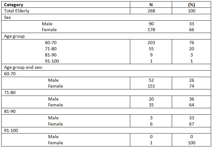
Based on the results of the profile data processing in the form of sex and age shown in Table 1 above, it is known that there are 268 elderly people consisting of 33% males (90 people) and 66% females (178 people), with 76% (203 people) in the age range of 60-70 years, 20% (55 people) in the age range of 71-80 years, 3% (9 people) in the age range of 81-90 years, and 1% (1 person) in the age range of 91-100 years. In this data, it is found that most of the elderly are female. In the study, stroke is more common in men aged 65-79 years although older women can also be at higher risk, namely those over 80 years 7,8.

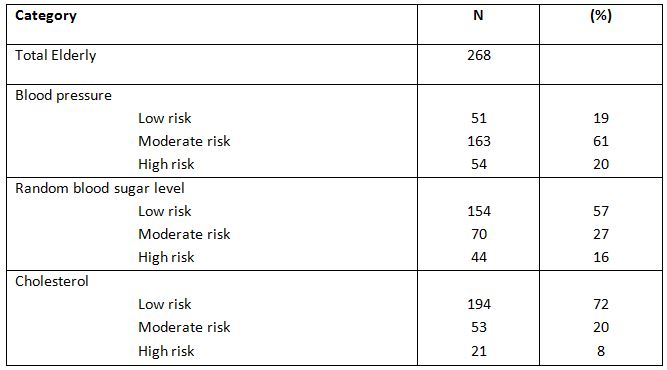
Based on the health profile shown in Table 3, there are three important factors that constitute indicators in the assessment of potential risks of stroke, such as:blood pressure, blood sugar, and cholesterol. A study also explains that those three indicators can be initial risk assessment for stroke conditions 5.

In the data processing table of the recorded results of medical examination based on blood pressure, it is concluded that 19% (51 people) of the elderly havea low risk potential, 61% (163 people) have a moderate risk potential, and 20% (54 people) have a high risk potential of stroke. It is also supported by a study conducted by Seshadri et al., which actually states that high blood pressure would result in higher risk of stroke in the elderly below 80 years old9. It is also visible from the data that such condition can also affect more than 70 percent of the elderly in the integrated health service center. A study conducted by Rodgers et al. also states that hypertension in the elderly can also result in a higher risk for the occurrence of stroke in the elderly5. A study conducted by Arboix et al. also states that high blood pressure conditions play a major role in increasing the risk of ischemic stroke in the elderly who are older than 85 years or more10.

In the data processing table of the recorded results of medical examination based on random blood sugar level,it was found that 57% (154 people) of the elderly have a low risk potential, 27% (70 people) have a moderate risk potential, and 16% (44 people) have a high risk potential of stroke. In general, the blood sugar condition and other conditions such as food management are also influential to the risk level of strokein the elderly 11.

In the data processing table of the recorded results of medical examination based on cholesterol, it was found that 72% (194 people) of the elderly have a low risk potential, 20% (53 people) have a moderate risk potential, and 8% (21 people) have a high risk potential. It can be said that most of the elderly are still safe in terms of cholesterol contained in the blood as a stroke risk parameter. Nevertheless, high cholesterol levels in the blood can be one of the risks that result in stroke among the elderly4. A study conducted by Reddy et al. stated that, besides the risks of diabetes and hypertension, dyslipidemia, obesity, smoking, and drinking alcohol can also increase the risk of stroke in the elderly12.

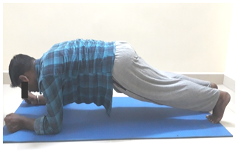
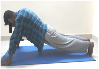
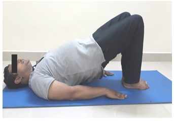
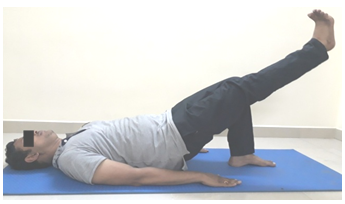
In this study, there were still more elderly women who came regularly, so that the data presented has not covered all the elderly. The data displayed is also only the data taken in the month of March. The activities in the integrated health service post for the elderly need to be enhanced with simple exercises that can increase physical activity in the elderly to help improve general health 13.

Ethical Clearance: Ethical clearance has obtained from Universitas Kristen Indonesia, Jakarta, Indonesia to conduct this study with reference number: 309/UKI.F8.D/PPM dated 01/06/2020.

Conflicts of Interest: The author declares that there is no competing interest in publishing this article.

Fund for the study: This is self-funded study.

Recommendation: The participation of elderly men in the integrated health service post needs to be increased, so that the elderly men who are at risk can be recorded because, based on data, many men have the risk of stroke. Health examination in the integrated public health service center for the elderly needs to be conducted regularly which needs to be supplemented with exercises or gymnastics for the elderly to improve their health condition. Simple exercises to increase physical activities can improve the health condition of the elderly.

CONCLUSION

As a conclusion, the elderly in the integrated health service center have quite various stroke risk potentials from moderate to high. In such conditions, information about health condition needs to be disseminated about the basic risks that would result in stroke also with initial handling for the prevention of stroke.

REFERENCES

  1. Hu, G., Sarti, C., Jousilahti, P., Peltonen, M., Qiao, Q., Antikainen, R., &Tuomilehto, J. (2005). The impact of history of hypertension and type-2 diabetes at baseline on the incidence of stroke and stroke mortality. Stroke, 36(12),2538-2543.
  2. Feigin, V. L., Krishnamurthi, R. V., Parmar, P., Norrving, B., Mensah, G. A., Bennett, D. A., & Davis, S. (2015). Update on the global burden of ischemic and hemorrhagic stroke in 1990-2013: the GBD 2013 study. Neuroepidemiology, 45(3), 161-176.
  3. Dávalos, A. (2005). Thrombolysis in acute ischemic stroke: successes, failures, and new hopes. Cerebrovascular Diseases, 20 (Suppl. 2), 135-139.
  4. Chen, R. L., Balami, J. S., Esiri, M. M., Chen, L. K., & Buchan, A. M. (2010). Ischemic stroke in the elderly: an overview of evidence. Nature Reviews Neurology, 6(5), 256-265.
  5. Rodgers, H., Greenaway, J., Davies, T., Wood, R., Steen, N., & Thomson, R. (2004). Risk factors for first-ever stroke in older people in the north East of England: a population-based study. Stroke, 35(1),7-11.
  6. Parr, E., Ferdinand, P., &Roffe, C. (2017). Management of Acute Stroke in the Older Person. Geriatrics, 2(3),27.
  7. Appelros, P., Stegmayr, B., &Terént, A. (2009). Sex differences in stroke epidemiology: a systematic review.  Stroke, 40(4), 1082-1090.
  8. Arboix, A., Oliveres, M., García-Eroles, L., Maragall, C., Massons, J., & Targa, C. (2001). Acute cerebrovascular disease in women. European neurology, 45(4), 199-205.
  9. Seshadri, S., Wolf, P. A., Beiser, A., Vasan, R. S., Wilson, P. W., Kase, C. S., & D’Agostino, R. B. (2001). Elevated midlife blood pressure increases stroke risk in elderly persons: the Framingham Study. Archives of internal medicine, 161(19), 2343-2350.
Citation:  Rosintan Milana Napitupulu, Novlinda Susy Anrianawaty Manurung (2021). Overview of the health profile of the elderly referring to the risks of stroke in the Sub-district of Cililitan, Jakarta, Indonesia , ijmaes; 7 (1); 927-932.

A study to find the effectiveness of iontophoresis with open kinematic chain exercises in pes anserine bursitis in sports persons

Gummadi Ashish1

Coresponding Author:

1Senior Physical therapist, Department of Neurorehabilitation, Institute of Neurosciences, Kolkata, Mail id: ashishgummadi@gmail.com

Aim and background of the study: Pes Anserine Bursitis is an inflammatory condition of pes anserine bursa which affects medial and inferio- medial aspect of the knee with more painful and swelling around the knee joint. Aim of the study is to find the effectiveness of iontophoresis and open kinematic chain exercises on pain, and knee range of motion of sports person with pes anserine bursitis.
Methodology: 30 male and female subjects according to inclusion and exclusion criteria were randomly divided into two groups for the study where, Experimental Group was treated with Iontophoresis and open kinematic chain exercise. Control Group was treated with Ultrasound therapy.
Result: The study reported  the Group  treated with Iontophoresis and open kinematic chain exercise got significant improvement in VAS and KROM score with p=0.001.
Conclusion: the study concluded that the experimental group which received iontophoresis along with open kinematic chain exercises demonstrated good effect in reducing pain and increasing Knee ROM.

Key Words: Iontophoresis, open kinematic chain exercises, Pes anserine bursitis, VAS, KROM.
Received on 14th January 2021, Revised on 22th January 2021, Accepted on 18th February 2021; DOI:10.36678/IJMAES.2021.V07I01.001

INTRODUCTION 

Knee is a hinge variety joint, which provides flexion and extension movements and a small degree of medial and lateral rotation. Pes anserine bursa of knee joint get inflamed due to over use and the bursitis affect medial and inferio- medial aspect of the knee with more painful and swelling around the knee joint 1, 2.

Pes Anserine Bursitis Epidemiology: Pes anserine bursitis is a common in running sports person. It is self-limiting condition, it can be cure with surgery or without surgery.1Pes anserine bursitis risk increases with endurance sports (distance running or triathletes), making changes to or beginning a new training program, sports that place stress on the muscles that insert at the pes anserine, such as those that require pivoting, cutting or, jumping, improper training, poor strength and flexibility, failure to warm-up properly before activity, improper knee alignment (knock knees), arthritis of the knees and trauma or constant friction on the bursa3.

Pes Anserine Bursitis in Sports Person (Marathon Runners): Pes Anserine bursitis risk is higher in endurance training and one of the most frequently affected individuals are Marathon Runners as they performs lots of endurance training. Usually in marathon runners, the tendon may become overused due to change in the training- for example running more uphill or just increasing the intensity of training program. Or any change in footwear can also be a cause 4.

 The new runners or those who have not had a professional gait analysis can develop Pes Anserine Bursitis. This Bursitis may develop over the course of long run, or may be a few weeks. Whatever be the duration, the main cause is due to overuse of the tendons that guards the pes anserine bursa.6

Pain in the inner aspect of knee, swelling and difficulty to bend the knee are the main symptoms of Pes Anserine Bursitis. Later stage muscle around the knee which produce extension and flexion movements may go for week followed by reduce the range of movement in knee joint 5.

Physiotherapy is the most widely used form of treatment adopted for gaining relief from pes anserine bursitis. It is used in both modes as single line of treatment including exercises or in form of combination with electrotherapy modalities like Ultrasound, Iontophoresis etc. The exercises include stretching, strengthening, ROM exercises. In flat feet individuals suggests with wearing arch supports in their shoes. Compression bandages or knee sleeves also helpful for reduce symptoms 6,7.

Iontophoresis is a therapeutic technique in which ion transfer into the body tissues by using electrical current as a driving force (LeDue, 1903).Iontophoresis is painless, sterile and non-invasive method to introduce specific ions into the body tissues. In case the ions are used in the form of ointment, a layer of its applied at the site to be treated. Iontophoresis treatment with dexamethasone is well tolerated by the majority of the patients5.

Open kinematic chain exercises (OKC) are the form of exercises in which the distal segment is mobile or not fixed like isolated joint exercise e.g. Seated leg extension. Open kinematic chain exercises are helpful to correct strength deficits of specific muscles or joints and beginning of rehabilitation when athlete not able to perform CKC exercises.11Open kinematic chain exercises are helpful to gain strength, flexibility and endurance of.3 hamstrings muscles group in sports person affected with Pes anserine bursitis8.

To evaluate the pain intensity VAS has been used whereas Knee ROM was measured using Goniometer. This study aimed to find the effectiveness of both the intervention in sports person with Pes Anserine bursitis.

In this modern age, there is a need of active research on an easy and effective intervention to reduce the pain and gain the near full ROM which enhances the performance in sports person.

Many Pes anserine bursitis patients experience pain, muscle weakness, decreased ROM and functional disability. Most patients regain their ROM muscle strength within a few week, but between 30 to 50 % are no longer able to get full ROM, muscle strength and functional ability after 1-2 month. So there is a strong need to gain full ROM muscle strength and pain reduction in Pes anserine bursitis rehabilitation .The persisting problem of ROM at knee muscles weakness and increased pain needs to be addressed and newer  strategies such as Iontophoresis with open kinematic chain exercises  can bring better insights for sports person with Pes anserine bursitis7,8.

In this study, iontohoresis with dexamethasone sodium and open kinematic chain exercise has been used to treat the sports person diagnosed with Pes Anserine bursitis.Iontophoresis is a therapeutic technique in which ion transfer into the body tissues by using electrical current as a driving force (LeDue, 1903).

Iontophoresis is painless, sterile and noninvasive method to introduce specific ions into the body tissues. In case the ions are used in the form of ointment, a layer of its applied at the site to be treated.11Iontophoresis treatment with dexamethasone is well tolerated by the majority of the patients and is effective in reducing symptoms such as pain and tenderness and better enable patients to tolerate therapeutic exercises and, therefore, will accelerate the rehabilitative process8.Hence this study is an attempt to find out the effectiveness of Iontophoresis with Open kinematic chain exercises (OKC)

METHODOLOGY

The subjects for the study were taken from the outpatient department of Neuro-rehabilitation Instuite of neurosciences kolkata. The subjects referred to the physiotherapy department with the diagnosis of Pes Anserine bursitis were screened for inclusion and exclusion criteria. The subjects who were qualified for the trial will be explained the aim and objectives of the study.

The subjects were requested to sign the consent form. The subjects were measured for their baseline outcome measures. A total 30 subjects who fulfil the inclusion criteria were included for the study and allotted into 2 groups, Experimental group (EG) and Control group (CG) randomly. The subjects with both groups received intervention for 12 days, the baseline measurements will be compared to the data at the end 12 days.

Procedure of intervention for the experimental group

Iontophoresis: Dexa-methasone sodium phosphate 0.4% with aqueous water was given via Galvanic current. The current used for this process was kept 1mA with subjects comfort considerations. In Iontophoresis current density always depends on quantity of current delivered per unit surface area. For this, two electrodes are used negative electrode (cathode) and positive electrode (anode), Negative electrodes used was double the size of the positive electrode.  Since Dexa-methasone sodium phosphate is negative ion (-), it was placed over the cathode (-). The cathode was placed over the pes anserine bursa where as the anode was placed over the calf muscle. Duration for iontophoresis was 20 minute per session once a day for 5-7 days 3

All the 15 patient of experimental group treated with Iontophoresis for initial 5 days were then treated with Open kinematic chain exercises (OKC) for knee everyday two session each session consisting  of 30 minutes .Each exercises was performed with proper rest period of  about 2 minutes after each exercises. Patient performed these exercises in supine lying position and support was provided by te assistant as necessary.

These exercises included the following exercises-

Hamstring stretch on wall: Subjects was in supine lying with hands by side, subjects involved leg on wall and other leg as free, as instructed by examiner, 10 repetitions were done with feel stretch.

Single leg hip extension: The patient was asked to lie down on his back with his knee bent at 90 degree and his feet hip width apart. From this position patient was asked to raise the leg and hold it suspended. Then the patient was asked to push his hips and lower back as one unit through the stabilizing leg, keeping his weight centered at his heel.10 repetitions with 10 counts hold

Seated knee extension: Subjects was in high sitting position, and the patient was asked to straight the knee.10 repetition with 10 count hold

Straight leg raise (SLR): The non-affected leg was flexed for 90 degree at the knee joint; the affected lower limb is straight on the table. The patient was asked to raise the affected lower limb straight to the level of the other leg.10 repetitions with 10 count hold.

Procedure of intervention for control group

Each participant in this group received ultrasound therapy with the intensity ranging from 0.8 -2 W/cm2 for 8 minutes in pulsed mode. The subject was positioned in high sitting position with the knee slightly rotated outward and the other leg of the subject was asked to keep apart from the involved limb.  The ultrasound was given by the therapist over the painful area in small concentric circles.33

Method for outcome measures

Visual Analogue Scale (VAS): VAS was used to measure subjects perception level of pain due to Pes anserine bursitis .It consists of scale where scoring is from 0-10. With score 0 subjects express no pain and with score 10 expresses maximum pain. The patient was asked to level the highest amount of pain they were getting at the time of assessment.

Knee Range of Motion (ROM)-Goniometry: Knee ROM includes flexion and extension which was measured by goniometer. To measure knee flexion, patient was asked to lie down in supine position and the subject was asked to bend the knee. Three trials of the same procedure were performed and average of the readings was considered for data analysis. To measure Knee extension, the patient was asked to lie down in prone position. The subjects was instructed to lift the knee without pelvic tilt (stabilize femur) and then goniometer was placed and the average reading of the three trials was recorded.

Inclusion criteria: Subjects between ages of 15-30 yrs. Both genders were included. History of improper training, running up hills with trackers, which produce pain and tenderness localized near Pes anserine bursa, Marathon runners, running for > 2 years were selected for the study.

Exclusion Criteria: Subjects with stress fracture, subjects with other lower limb musculoskeletal pathology, subjects with ischemic disorder of lower limb, subjects with muscle spasm of hip extensors, malignancies or tissue damaged by radiation therapy, subjects with visual impairment   or  with  any   other  neurological

disability like any head trauma, dementia, learning disorder and schizophrenia, and Osteoarthritis of knee were excluded from the study.

Materials and Methods: It includedCouch, EMS/Galvanic, Dexamethasone sodium phosphate aqueous solution, Cotton, Microspore tape, Goniometer, Ultrasound machine, and Aquous gel

Outcome Measures: Visual analogue scale (VAS) and Knee range of motion (ROM) goniometry

RESULT

Study design: A Comparative two group interventional clinical study

Table 1: Age distribution of patients studied, P=0.909, Not Significant, Student t test

Table 1 show that there is 46.7% of patient in Experimental group and 40% in Control group of age group 15-20. However, 53.3% were in

experimental group and 60% in control group of age group 21-30. 

Table 2: Gender distribution of patients studied, P=0.713, Not Significant, Chi-Square Test

In table 2, Experimental group consists of 60% female and 40% male whereas control group consists of 53.3% female and 46.7% male patients. Overall, 56.7% of patient was females and 43.3 % was males.

Table 3: VAS Score-A Comparative assessment at pre and post in two groups of patients studied

VAS score of both the group were analysed in table 3 and the mean difference in percentage was obtained. Range of VAS scale score was made as 0, 1-3, 4-6, 7-10 in both the groups. The VAS score has been significantly reduced in Experimental Group as compared to Control Group. In the VAS score the % difference between the pre and post treatment is 46.7%, 53.3%, 0% and -100%.

Table 4: Knee ROM- A Comparative assessment at pre and post in two groups of patients studied

In table 4, Knee ROM has been ranged as 50-80, 80-100,100-120,120-135 degree. The % difference between pre and post treatment is -73.3%,-26.7%, 66.7%, and 33.3% which shows the high significance of the treatment in Experimental Group. The P value is of <0.001** resulting highly significant. In control Group, the KROM in day 14 is 60%, 33.3%, 6.7%, 0% respectively for the given ROM range which indicates that there is very less improvement in KROM  after treatment. The mean difference is not as high as that of experimental Group which is -13%, 6.6%, 6.7% and 0.0% respectively for the given range.

Table 5: A Comparative assessment of VAS and Knee ROM at pre and post in two groups of patients studied

In table 5, VAS score of the patient in EC was 8.27±0.80 whereas post treatment in day 14 it became 0.67±0.72. The mean difference noted is 7.600 and the p value is highly significant at <0.001**. Similarly for control group, VAS score in day 1 is 7.80±0.94 and for day 14 is 4.40±0.83. There is not much difference between pre and post score for VAS in control group, which is just 3.400.

Knee ROM for EC in day 1 was 69.67±14.82 whereas in day 14 it is 119.67±6.67.The mean difference between pre and post treatment score is of -50.000 and the P value at <0.001**. However, control group doesn’t depict much change after treatment. The mean difference between Pre and Post treatment score is just -13.333, far less than that we achieved in EC.

Table 6: Comparison of difference of VAS and Knee ROM in two groups of patients studied

Table 6 displays the difference in VAS and Knee ROM of patients of. Experimental group and control group. Difference in VAS score is 5.50±2.42 and Knee ROM is 31.67±22.18.

DISCUSSION

The purpose of the study was to find the effectiveness of iontophoresis with open kinematic chain exercises to reduce pain and to increase knee ROM in sports person with pes anserine bursitis. The study consists of 30 sports persons diagnosed with Pes Anserine bursitis.15 subjects were kept in experimental group and other 15 in control group. The subjects in experimental group received iontophoresis with open kinematic chain exercises of knee and the control group was treated with ultrasound only. In this study, experimental group and control group result were analysed using student t test and chi square test.

Result obtained from this present study shows that when the mean reduction values of VAS and ROM were analyzed within the groups, it was statistically significant in both the groups for VAS score whereas there was no any effect in KROM in control group. Iontophoresis along with open kinematic chain exercises was found to be very effective in reducing pain and improving ROM. VAS score assessment for pain showed significant improvement in both the groups after the treatment.

This statement is supported by Robert et al (2003) where they concluded that iontophoresis administration of Dexa-methasone sodium phosphate is well tolerated by the patients and is able to reduce pain.The statement is further supported by Linda C et al (1995) where dexa-methasone sodium phosphate was used to treat rheumatic arthritis and the result obtained showed that iontophoresis treatment was more effective in reducing pain and improving ROM. The dexamethasone sodium phosphate delivered via iontophoresis penetrates deep into the joint and thus reduces the symptoms 9.

Iontophoresis is the most commonly used method for pain and inflammation. Iontophoresis for treatment of locomotor system lesions should be considered as an alternative to peri and intra-articular injections because the drugs used diffuse to the tissues along the dermal barrier and penetrate into the body and thereby reduces the symptoms more effectively.Iontophoresis administration of dexa-methasone sodium phosphate was more effective in reducing the pain and other symptoms in short term follow up for patients with lateral Epicondylitis.

Although ultrasound was found to be reducing pain and increase the healing process, the result obtained from this study was not as highly significant as in experimental group. Therefore all the above literature supports the efficacy of iontophoresis in reducing the pain and allowing the better tolerance for exercises.The study was designed to find the effectiveness of iontophresis and open kinematic chain exercises   to reduce pain and to improve KROM in sports person with Pes anserine Bursitis.

This study was a randomised experimental study where 30 sports person with Pes anserine bursitis of age group 15-30 wereselected.The subjects were evaluated and included for the study using inclusion criteria such as VAS score 6-9, both the gender and tenderness over the pes anserine bursa. Subject with recent surgery in and around the knee, stress fracture, and other progressive neurological deficit were excluded. Subjects who were willing to participate in the study were requested to fill the consent form.

On day one, pre- treatment baseline assessment was done using VAS for pain assessment and knee ROM by using goniometer.30 Subjects were assigned to either group of 15 in each group. That is experimental group (EG) which received iontophoresis and OKC exercises and control group (CG) which received ultrasound therapy. Post treatment assessment was done on 12th day of treatment using VAS and by measuring KROM using goniometer. Data were analysed using Paired –“t” test and Un-paired “t” test. The result shows that there is reduction of pain and improvement in KROM after 12 days of intervention in EG when it is compared to CG.

Therefore the study concludes that the iontophoresis and open kinematic chain exercises are an effective intervention to reduce pain and to increase KROM in sports person with Pes Anserine bursitis after the 12 days of treatment. The analysis found the highly significant differences in between the pre and post test score of KROM for experimental group. This statement is supported by M Seshagirirao et al (2016) which concluded that open kinematic chain exercises are effective in improving the strength and knee function 10.

Graham et al (1993)found that Open kinematic chain exercises are helpful in improving range of motion and strength of quadriceps and hamstrings muscles. This statement is further supported by the study William E Prentice (2003) who observed that open kinematic chain exercises are helpful to gain strength, flexibility and endurance of hamstrings muscles group in sports person affected with Pes Anserine bursitis.  Open kinematic chain exercises of knee were studied and the result obtained showed significant improvement in the hamstrings strength and ROM. Therefore all the above literature supports the efficacy of iontophoresis and open kinematic chain exercises in reducing pain and improving knee ROM in sports person with Pes anserine bursitis11.

As Pes anserine bursitis is considered as a self- limiting disorder, in some cases it is not possible to determine if this self-limiting factor led to the improvement of pain levels and knee ROM instead of treatment administered. The sample size is small and the treatment duration is for short period of time. In future, further studies can be done on larger sample size and for long duration. Additionally, further studies can be done which involves treatment programs like iontophoresis and close kinematic chain exercises.

Ethical Clearance: Ethical clearance has obtained from Institute of Neuroscience, Kolkata to conduct this study with reference number: 214/ Research/IRB/2018-19dated 24/09/2019.

Conflicts of Interest: The author declares that there is no competing interest in publishing this article.

Fund for the study: This is self-funded study.

CONCLUSION

This study concluded that the experimental group which received iontophoresis along with open kinematic chain exercises demonstrated   good effect in reducing pain and increasing Knee ROM.  But when the experimental groups is compared with the control group which received only ultrasound therapy, EG showed significant improvement in VAS and KROM score whereas there is not much significant improvement in outcome measures post treatment in control group.

REFERENCE

  1. Miller RH III. (1998)Knee injuries. In: Canale ST, ed. Campbell s operative orthopaedics. St Louis: Mosby; 1113–1299. .
  2. Pompan DC. (2016). Pes Anserine Bursitis: An Underdiagnosed Cause of Knee Pain in Overweight Women. Am Fam Physician.  Feb 01; 93(3):170. 
  3. MSeshagiriRao et al. (2016). Effectiveness of open kinematic chain exercises versus closed kinematic chain exercises of knee in meniscal instability athletics. International Journal of Physiotherapy and Research 4(1):1345-1351 · 
  4. McMorrisy and perry et al, (2005). Effect of open vs close kinematic chain exercises in extensor resistance training in knee injury patient.
  5. Sim J, Waterfield J. Validity, (1997). Reliability and responsiveness in the assessment of pain. Physiotherapy theory and Practice; 13: 23-27.
  6. Revill S, Robinson J, Ronsen M and Hogg M. (1976).The reliability of a linear analogue for evaluating pain.Anaes; 31:1191-1198.
  7. Gogia PP, Braatz JH, Rose SJ, Norton BJ. (1987). Reliability and validity of goniometric measurements at the knee.  67(2):192-5.
  8. Escamilla RF,Fleising GS, Zheng N, Barrentine SW, Wilk KE, Andrews J R. (1998). Medsci sports exercises apr 30; (4).
  9. Lysholm J, Gillquist J. (1982). Evaluation of knee ligament surgery results with special emphasis on use of a scoring scale. Am J Sports Med 3; (10): 150-4.
  10. Neeter C, Thomee R, Silbernager KG, et al. (2003). Iontophoresis with or without dexamethasone in the treatment of acutachilles tendon pain. Scand J Med Sci Sports; 3:376-382.
Citation:   Gummadi Ashish (2021). A study to find the effectiveness of iontophoresis with open kinematic chain exercises in Pes Anserine bursitis in sports persons, ijmaes; 7 (1); 916-926.

A brief review in non-specific low back pain: Evaluation and physiotherapy intervention

Lucky Anggiat1*

Author:

1Physiotherapy Program, Fakultas Vokasi, Universitas Kristen Indonesia, Indonesia

Corresponding  Author :

*1Physiotherapy Program, Fakultas Vokasi, Universitas Kristen Indonesia, Indonesia

Mail id: lucky.panjaitan@uki.ac.id

ABSTRACT

Background: Low back pain was the most common case in musculoskeletal disorders. Non-specific low back pain (NSLBP) described as low back pain with no clear causal relationship between the symptoms, physical findings, and imaging findings. This study aimed to briefly review the evidence the evaluation and intervention for NSLBP in physiotherapy practice.
Methods: In this study, the library research method was used, which took online and offline data sources referring to books, journals, articles related to the topic of evaluation and physiotherapy intervention in NSLBP conditions as a data source to answer research objective
Results: Some researchers commonly assess the pain, range of motion (ROM), functional ability and quality of life. As a regular treatment for non-specific low back pain, some study recommended general therapeutic exercise and manual therapy to reduce the problematic of non-specific low back pain.
Conclusion: Physiotherapist can evaluate patients with NSLBP based on the patients complains like pain using VAS or NPRS, Lumbar ROM using schober method, functional disability using ODI or RMDQ and Quality of life. For the physiotherapy intervention of non-specific low back pain, we can summarize that the first-line management of NSLBP is self-management exercise. Furthermore, physiotherapist can use any method of therapeutic exercise and manual therapy to reduce pain, improve lumbar ROM, increase functional ability and quality of life.  

Keywords: Non-specific Low Back Pain, Evaluation, Intervention, Physiotherapy

Received on 08th August 2020, Revised on 22nd August  2020, Accepted on 29th August 2020, DOI:10.36678/IJMAES.2020.V06I03.001

INTRODUCTION

Low back pain was the most common case in musculoskeletal disorders 1. The challenge when managing the low back pain was diagnosing the low back pain and choose the treatment 2. In general, the low back pain consists of two types, which is specific and non-specific low back pain (NSLBP). Low back pain with specific type can be divided into low back pain that related to vertebrae and non-vertebrae. In the other hand, NSLBP described as low back pain with no clear causal relationship between the symptoms, physical findings, and imaging findings 3. In addition, NSLBP is classified into low back pain, which is not related to the neurological problem and degenerative syndrome 4.

Previously, some research showed that NSLBP mainly affected the young population. Non-specific low back pain has also increased in the general community, which have affected the adolescents and middle-aged worker 5. Nordin et al. reported that the incidence of NSLBP among undergraduate student in health sciences programs was 40.3%, the incidence was associated with the age, years of study, physical fitness, and hours they spent sitting in the classroom 6. Another research by Anggiat et al. showed a similar result that students in a university experience NSLBP with 74.6% 7. Also, a community-based program revealed that teachers also experience low back pain with more than half of the teacher population 8.

Numerous research already conducted to evaluate the non-specific low back pain. The most common evaluations were pain assessment, trunk flexibility, functional ability and quality of life 9,10,11. Those evaluations seem to be correlated with the population which experienced NSLBP because the NSLBP mainly causes pain, reduced flexibility of trunk movement, functional limitation and sometimes also affected to quality of life 12. Furthermore, most studies also conducted in the physiotherapy intervention for NSLBP. Physiotherapy was the first line care to give intervention in person who experienced NSLBP 13. Some research was done resulting recommendation of using home exercise program, manual therapy and general exercise therapy 2, 14, 15.

From the background, this study aimed to briefly review the evidence the evaluation and intervention for NSLBP in physiotherapy practice. In addition, with this article, the physiotherapist will easily use the evidence to treat the non-specific low back pain.

METHOD

In this study, the library research method was used, which took online and offline data sources referring to books, journals, articles related to the topic of evaluation and physiotherapy intervention in NSLBP conditions to answer research objective. The use of books is focused on basic science such as evaluation and applied anatomy. In the intervention, articles in journals and clinical guidelines were used that recommend physiotherapy in NSLBP conditions. The searching strategy was conducted a literature search using the online database in google scholar for ‘‘physiotherapy for non-specific low back pain’’ The bibliographies of particularly relevant articles were searched as well.

RESULT AND DISCUSSION

Applied anatomy and contributing factor of NSLBP: Non-specific low back pain mainly related with posture or poor body mechanic. There are several other factors caused the NSLBP with anatomical problem. Some anatomical related factors can be contributed to the incidence of NSLBP. Lumbar flexion motion is performed by rectus abdominis, external and internal oblique. For extension motion is performed by illiocostalis, longissimus, semi spinalis, multifidus. For lateral flexion motion, performed by quardatus lumborum and also assisted by illiocostalis, longissimus, spinalis. For rotation motion, the prime mover is tranversus abdominis, and multifidus work contralateral for every rotation motion 16. Some muscles of the back that related with the low back pain are tranverse abdominis, internal obliques, erector spinae and multifidus. Those muscles will indicate low back pain problem if the muscle has poor muscular endurance, which is related with, prolonged posture activity 17. Currently, multifidus muscle dysfunction is being implicated as a contributory factor in the development or recurrence of sub-acute and chronic back pain 18.

In general, NSLBP has two classifications; an acute stage, which pain lasts less than 12 weeks and chronic stage, which pain lasts more than 12 weeks 19. According to Taguchi, chronic NSLBP was due to physiological structural fragility in lumbar region, and often caused by improper posture, which can be called a living functioning impairment 4. A research showed that mobility of the spine reduced causing disorders in muscle synergies and consequently increased the energy cost of maintaining postural ability 14.

One of the causes of NSLBP is postural pain caused by prolonged sitting activity for about 2 hours a day, which causes increased discomfort in the body 20,21. Sitting in a slumped position is also associated with fatigue in the internal oblique muscles and/or transverse abdominis which keeps the spine in one position so that it makes these muscles prone to injury and can also be caused by atrophy of the multifidus and para-spinal muscle 21.

Another study also reported that sitting in a position such as the excessive kyphotic posture with frequent lumbar flexion or the excessive lordotic posture with too much extension can result in low back pain 22. A study in Japan, it was reported that 22% of the population aged 20-85 years had NSLBP 23. In line with several other studies, it also reported that students, office workers and teachers also experience NSLBP due to prolonged sitting for at least 3 hours a day 6,7,8. The pain produced by NSLBP in a population of both students, teachers and office workers can affect physiological and psychological stress and sometimes cause secondary disturbances in the form of decreased quality of life 4, 5.

Evaluation for NSLBP: To assess the effect of low back pain to individual life, the physiotherapist will carry out several assessments to evaluate low back pain and its impact. Some researchers commonly assess the pain, range of motion (ROM), functional ability and quality of life 9,10,15,24. Physiotherapist commonly uses the visual analogue scale (VAS) to assess the pain perception of low back pain 15, 25. In a study by Hawker et al. shouted that the VAS is self-completed by the respondent.

They also mentioned that the VAS is widely used due to simplicity and adaptability to a broad range of population and settings 26. As a subjective measurement of pain, VAS consists of a 10 cm line with two end-points representing ‘no pain’ and ‘worst imaginable pain’. Patients will have asked to rate their pain by placing a mark on the line corresponding to their current level of pain 27.

The VAS is a well-known assessment tool for pain and recommended as a means of rating the subjective pain 20. Another version with the same meaning of VAS, physiotherapist can use Numeric Pain Rating Scale (NPRS) which consist of number 0 for no pain until 10 which worst worst imaginable pain. Physiotherapists can use either of these pain evaluations because those pain evaluation tool have the same validity and reliability 26.

As the pain was assessed in the non-specific low back pain, another assessment that related with pain was the flexibility of the lumbar spine, where the pain will affect the flexibility of lumbar spine 28. Furthermore, a study by Wong and Lee describes that there is a correlation between patients with LBP and the decreased lumbar ROM 29. The flexibility of the lumbar spine is related to the lumbar range of motion (ROM). They also conclude that the lumbar ROM should be evaluated after the treatment of LBP to know the effects of the treatment. Previously, some researcher decided to use a measuring tape to evaluate the lumbar ROM 10,30. Tape measurements were the least expensive method to measure spinal movement and perhaps the easiest to use 31. In order, to measure flexion and extension of lumbar, the modified Schober method can be used by the physiotherapist 32. Some studies also reported that the Schober method was one of the good methods to assess the lumbar flexibility 32, 33.

In order, to measure lateral flexion, the fingertip-to-floor method can be used as an additional measurement of lumbar ROM 31. However, flexion and extension were the most commonly used as the main evaluation of the lumbar range of motion in low back pain cases 10, 34.Clearly, the flexion and extension movement is the main segmental movement of the lumbar spine 35.

The risk of people with NSLBP who have high pain score may also develop functional disability 36. Furthermore, to evaluate disability, should to use disability measurement along with pain measurement. In other studies, it was reported that pain was also associated with impaired functional activity, which in this case could cause disability, where a high rate of disability was associated with high pain rates 37. Activity disorders that can occur in patients with NSLBP are activities indoor and outdoor activity such as on travelling, climbing and descending stairs, walking, wearing clothes, eating, using the toilet, using public transportation and other social activities 38.

Several studies provide recommendations using the Oswestry Disability Index (ODI) for assessment of functional disability caused by LBP 39,40,41. The Oswestry disability index (ODI) is aimed specifically at LBP conditions and is the best standard in evaluating the functional activity of people with NSLBP both before and after the intervention 39. Furthermore, research from Fairbank and Pynsent stated that ODI has been translated into several languages including English and can then be used validly and reliably for examining conditions of back pain related to disability evaluation 42.

On the other hand, other evaluation related to functional activities, the physiotherapist can use the Rolland-Morris Disability Questionnaire (RMDQ) 43. In general, RMDQ also has similar validity and reliability to ODI, but in detail, ODI is still superior in evaluating NSLBP patients 44, 45.

Furthermore, the RMDQ is still considered a very useful evaluation tool in evaluating the condition of NSLBP patients. Similar to ODI, RMDQ also evaluates the patient’s status in terms of pain, dysfunction and disability 43. Thus, evaluation before and after the intervention can be carried out using the questionnaire to see the changes that occur before and after the intervention.

Non-specific low back pain with a long period of time can affect the quality of life. Pain that is quite severe has contributed to reducing the quality of life of an individual 46. Furthermore, in their study, it was concluded that the LBP condition was quite severe and reduced activity was also associated with physical functional disorders and even caused mental disorders and individual productivity.

Followed by other research by Tsuji et al. also reported that the condition of pain was quite high and disturbances in quality of life affected the decrease in productivity of workers with NSLBP 47. Furthermore, they also suggested to be able to evaluate the quality of life of individuals who have NSLBP as an essential examination. In field of education, Kennedy et al. also reported that students with LBP affect their quality of life by decreasing psychosocial aspects such as experiencing sadness, being overwhelmed and exhausted 48.

Evaluation of quality of life generally uses the Health-related quality of life (HRQoL) questionnaire. The questionnaire in the quality of life examination is generally classified into generic, condition specific, or patient specific. In evaluating the HRQoL in low back pain population, the condition-specific instrument of HRQoL can be used.

The HRQoL is a multidimensional concept that refers to function and well-being on various dimensions of health, including physical, emotional, social and spiritual aspects of life 49. Some of the HRQoL instruments that can be used to assess the quality of life on low back pain patient is the Short Form 36-items Health Survey (SF-36) or with other short versions such as SF-12 or SF-8 50.

Physiotherapy Intervention: As a regular treatment for non-specific low back pain, some study recommended general therapeutic exercise to reduce the impairment of NSLBP  51, 52. The type of general exercise for low back pain mainly is an active stretching, which easily performed by the patient 53. A study by Gawda et al. revealed that the stretching therapy that done by the patients could be effective to reduce the low back pain 14. In their study, the physiotherapist gives some example to do the stretching until the patients can do the stretching by themselves. Some guidelines, also reported some educational exercise that can be done by the low back pain patient to manage the low back pain 19,54. In 2016, National Institute for Health and Care Excellence (NICE) produce an assessment and management guideline for the low back pain. In that guidelines, described that the self-management was the first management for low back pain 13.

A review study by Bardin, King and Maher also recommended self-management exercise along with hot-pack as the first line care for NSLBP 2. The use of hot packs considered as a pain relief that provides analgesia effect and muscle relaxation 2,55. Self-management exercise or educational home exercise program with hot packs also recommended by a health care guideline as a management of low back pain patient 54. A research by Taguchi stated that the therapeutic heating is often conducted by physiotherapy for the chronic NSLBP, despite the effectiveness in not clear, however, from the viewpoint of relaxation, the purpose of therapeutic heating is reducing the pain 4.

Furthermore, several clinical guidelines provide recommendations for interventions in the management of LBP in clinical practice. In America, the clinical practice guidelines made by the American Physical Therapy Association (APTA) recommend several interventions that can be used in general in LBP patients 43.

The first recommendation is to use manual therapy with joint mobilization or thrust manipulation to reduce pain and disability. Then, for therapeutic exercise intervention is recommended using back-specific strengthening, coordination and endurance exercises. Those two recommendations are based on strong recommendations. In addition, it is also recommended to use patient education/counseling for disruption of activities due to LBP and increase endurance with fitness and endurance activity based on moderate and strong recommendations.

Another clinical recommendation in the United Kingdom also provides several options in physiotherapy intervention in LBP conditions 13. In addition to using self-management exercises, physiotherapists are also recommended to use exercise therapy such as exercises based on biomechanics, aerobics or in combination with other types of exercise. Just like in APTA guidelines, in the next recommendation, physiotherapy is also recommended using manual therapy such as mobilization or spinal manipulation and also soft tissue manipulation. Besides, they also recommend using psychological therapy such as the cognitive-behavioral approach in combination with exercise therapy or manual therapy.

In a study by Oliviera et al. that reviewed clinical practice guidelines for the management of NSLBP, recommended several suggestions related to physiotherapy interventions in NSLBP 56. In general, in the types of physiotherapy interventions, they recommend exercise therapy for the management of both acute and chronic NSLBP. Although the types of exercise therapy are still considered inconsistent, physiotherapists can use various types of exercise therapy according to the therapist’s ability. Subsequently, manual therapy with spinal manipulation has also become a recommended intervention in the management of both acute and chronic NSLBP.

In manual therapy, the physiotherapist can choose to use spinal mobilization or manipulation, however, exercise therapy has several types of methods that can be used according to the physiotherapist’s ability to perform the methods 13,43. Exercise therapy that can be recommended is the motor control exercise approach and core stability exercises 57,58. Exercises using a flexion and extension approach as known as the directional preference of McKenzie method or the other name is Mechanical Diagnosis and Therapy (MDT) can be used as well 59.

Furthermore, exercise therapy with a proprioceptive approach such as Proprioceptive Neuromuscular Facilitation which is rarely used in musculoskeletal cases, can be used in NSLBP patients as well. All types of exercise therapy have a beneficial effect in reducing pain, increasing lumbar ROM and improving functional activity and quality of life with no superiority among each other methods 41,60.

This study is a simple short review study in the evaluation and intervention of physiotherapy in NSLBP conditions. There are still many limitations in this research, so that, in the future, a more comprehensive review study method should be carried out.

CONCLUSIONS

Numerous research has been able to provide very useful information to the physiotherapist to evaluate and treat the patients with NSLBP. Based in this present study, we can conclude that physiotherapist can evaluate patient with NSLBP based on the patient’s complaints such as pain using VAS or NPRS, Lumbar ROM, functional disability using ODI or RMDQ and quality of life questionnaire.

For the physiotherapy intervention of NSLBP, we can summarize that the first-line management of NSLBP is self-management exercise. Furthermore, physiotherapist can use any method of therapeutic exercise and manual therapy to reduce pain, improve lumbar ROM, increase functional ability and improve the quality of life.

Conflict of interest: The author has no conflict of interest to declare.

Funding of study: This study has no funding or sponsorship was received. It is self-financed study.

Compliance with Ethics: This study based on review of previous conducted studies with does not contain any studies with human or animals.

REFERENCES

  1. Maher, C., Underwood, M., Butchbindee, R. (2017). Non-specific low back pain. Seminar, Lancet 389: 736-47
  2. Bardin L, D., King P., Maher C, D. (2017). Diagnostic triage for low back pain    : a practical approach for primary care. The Medical Journal Australia 206 (6), 268-273.
  3. Balagué, F., Mannion, A. F., Pellisé, F., & Cedraschi, C. (2012). Non-specific low back pain. The lancet, 379(9814), 482-491.
  4. Taguchi, T. (2003). Low back pain in young and middle-aged people. Journal of Japan Medical Association. JMAJ, 46(10): 417-423.
  5. Aziz, S., Ilyas, S., Imran S., Yamin F., Zakir A., Rehman A., Adnan S., Khanzada S. (2016). Effectiveness of McKenzie Exercise in Reducing Neck and Back Pain Among Madrassa Student. International Journal Physiotherapy 3 (1), 78-85.
  6. Nordin, N. A., Devinder, A. S., & Kanglun, L. (2014). Low Back Pain and Associated Risk Factors among Health Science Undergraduates. Sains Malaysiana, 43(3), 423–428.
  7. Anggiat, L., Hon, W. H. C., & Baait, S. N. (2018). The incidence of low back pain among university students. Pro-Life, 5(3), 677-687.
  8. Budhyanti, W., Anggiat, L., & Juwita, C. P. (2020). Pencegahan dan penanganan nyeri leher dan punggung bawah dengan peregangan mandiri pada pegawai sekolah menengah kejuruan negeri 10 cawang. Jurnal Fisioterapi dan Rehabilitasi,  4(1), 79-89.
  9. Dhaliwal, M. K., Amandeep, Jagmohan., Manjeet. (2014). To compare the effect of proprioceptive neuromuscular facilitation program versus core stabilization exercise for decreasing pain and improving function in patients with low back pain. IOSR Journal of sport and physical education 1. PP 29-35.
  10. Park, K.Y., Seo, K.Y. (2014). The Effects on the pain index and lumbar flexibility of obese patients with low back pain after PNF Scapular and PNF pelvic patterns, Journal of Physical Therapy Scienes 26 : 1571-1574.
  11. Anggiat, L., Hon, W. H. C., & Sokran, S.N.B.B.M. (2018). Comparative effect of proprioceptive neuromuscular facilitation and mckenzie method on pain in non-specific low back pain among university population. International Journal of Medical and Exercise Science, 4(3), 475-485.
  12. Pergolizzi, J. V., & LeQuang, J. A. (2020). Rehabilitation for Low Back Pain: A Narrative Review for Managing Pain and Improving Function in Acute and Chronic Conditions. Pain and Therapy, 1-14.
  13. National Institute for Health and Care Excellence (NICE). (2016). Low back pain and sciatica in over 16s: assessment and management. United Kingdom : NICE Guidelines. nice.org.uk.
  14. Gawda P, Dmoszyńska-Graniczka M, Pawlak H, Cybulski M, Kiełbus M, Majcher P, Buczaj A, Buczaj M. (2015). Evaluation of influence of stretching therapy and ergonomic factors on postural control in patients with chronic non-specific low back pain. Annals of Agricultural and Environmental Medicine 22(1): 142-146.
  15. Waqqar, S,. Rehman S, S., Ahmad, S. (2016). McKenzie treatment versus mulligan sustained natural apophyseal glides for chronic mechanical low back pain. Pakistan Journal of Medical Sciences 32(2):476-479.
  16.  Cael, C. (2010). Functional Anatomy: musculoskeletal anatomy, kinesiology, and palpation for manual therapists. Lippincott Williams & Wilkins, a Wolters Kluwer business. Philadelphia, PA.
  17. Kisner, C., Colby, L.A. (2012). Therapeutic Exercise: Foundation and techniques 6th edition, F.A. Davis Company. USA
  18. Danneels, L., Dickx, N and Cagnie, B.(2010) Low Back and Pelvic Pain, 7th Interdisciplinary world congress, Proceedings. p.186-195.
  19. Hussein A.M.M., Singh D., Mansor, M., Kamil, O.I.M., Choy, C.Y. Vijayan, R.(2009). Malaysian Low Back Pain guideline. First Edition. Malaysian Association for the Study of pain and Spine society Malaysia
  20. Casser HR, Seddigh S, Rauschmann M. (2016). Acute lumbar back pain-investigation, differential diagnosis and treatment. Deutsches Ärzteblatt International, 113: 223-34.
  21. Waongenngarm, P., Rajaratnam, B. S., & Janwantanakul, P. (2016). Internal Oblique and Transversus Abdominis Muscle Fatigue Induced by Slumped Sitting Posture after 1 Hour of Sitting in Office Workers. Safety and Health at Work 7(1): 49-54.
  22. Dankaerts W., O’Sullivan P., Burnett, A., Straker, L. (2006). Differences in sitting posture are associated with nonspecific chronic low back pain disorders when patients are subclassified. SPINE 31 (6), pp 698-704.
  23. Suzuki H, Kanchiku T, Imajo Y, Yoshida Y, Nishida N, Taguchi T. (2016). Diagnosis and Characters of Non-Specific Low Back Pain in Japan: The Yamaguchi Low Back Pain Study. PLoS ONE 11 (8): e0160454.
  24. Longo G, Loppini M, Denaro L, Maffulli N, Denaro V. (2010). Rating scales for low back pain. British Medical Bulletin, 98 ; 81-144.
  25. Hosseinifar M., Akbari M., Behtash H., Amiri M., Sarrafzadeh, J. (2013). A Comparison between the Effects of Stabilization and Mckenzie’s Exercises on the Pain, Disability, and Lumbo-Pelvic Stability in Patients with Non-Specific Chronic Low Back Pain” Life Sciences Journal ,10(10s); 298-302.
  26. Hawker G.A., Mian, S., Kendzerka, T., French, M. (2011). Measures of Adult Pain. American College of Rheumatology. Arthritis Care & Research 63 (11): pp S240–S252,.
  27. Huskisson EC. (1974). Measurement of pain. Lancet, 2(7889):1127-31.
  28. Lee A, S., Cholewicki, J., Reeves N, P, Zazulak B,T., Mysliwiec, L, W. (2010). Comparison of Trunk Proprioception Between Patients With Low Back Pain and Healthy Controls. Archives of Physical Medicine and Rehabilitation 91 (9):1327-1331.
  29. Wong, T, K, T and Lee, R, Y, W. (2004). Effect of low back pain on the relationship between the movements of lumbar spine and hip. Human Movement Science 23 : 21-34. doi:10.1016/j.humov.2004.03.004.
  30. Franklin, C.V.J., Kalirathinam, D., Palekar T., Nathani N. (2013). Effectiveness of PNF Training for Cronic Low Back Pain. IOSR Journal of Nursing and Health Science (IOSR-JNHS) 2 :41-52.
  31. Reese, N., Bandy, W. (2002). Joint Range of Motion and Muscle Length Testing. Saunders Company. Philadelphia, USA.
  32. Sihawong R., Sitthipornvorakul, E., Paksaichol, A., Janwantanakul P. (2016). Predictors for chronic neck and low back pain in office workers: a 1-year prospective cohort study. Journal of Occupational Health 58: 6-24.
  33. Chhaya, V., Renuka D., Vijaya K., Amol B. (2015). Lumbar range of motion : Reliability between schober’s test and modified schober’s test. Romanian Journal of Physical Therapy 21/ Issue 35/ may/ pp
  34. Kumar A., Zutshi K,. Narang N.(2011). Efficiacy of Trunk Propioceptive Neuromuscular Facilitation Training on Chronic Low Back Pain. International Journal of Sports Science and Engineering 5 (3): pp. 174-180.
  35. Key, J., Chaitow L. (2010). Back Pain: A Movement Problem, A Clinical Approach incorporating relevant research and practice. 1st Edition, Churchill Livingstone, Elsevier. UK.
  36. Melton, B, L., Moqbel, M., Kanaan, S., Sharma, N, K. (2016). Structural Equation Model of Disability in Low Back Pain, Spine, 41(20), 1621.
  37. Davis, D, S., Mancinelli, C, A, Petronis, J, J., Bensenhaver, C., Mcclintic, T., Nelson, G. (2013). Variables associated with level of disability in working individuals with nonacute low back pain: a cross-sectional investigation. Journal of orthopaedic & sports physical therapy 43 (2), 97-104.
  38. Di Iorio, A,.Abate, M,. Guralnik, J,M., Bandinelli,S,. Cecchi, F,.Cherubini,A., Corsonello, A.,  Foschini, N., Guglielmi, M., Lauretani, F., Volpato, S., Abate, G., Ferrucci, L. (2007). From Chronic Low Back Pain to Disability, a Multifactorial. Spine (Phila Pa 1976). 32(26): E809–E815.
  39. Seif, H.E., et al. (2015) The Effect of Stretching Hamstring, Gastrocnemius, Iliopsoas and Back Muscles on Pain and Functional Activities in Patients with Chronic Low Back Pain: A Randomized Clinical Trial. Open Journal of Therapy and Rehabilitation 3: 139-145.
  40. Vincent, J.I., MacDermit, J.C., Grewal, R., Sekar, V.P., Balachadran, D. (2014). Translation of Oswestry Disability Index into Tamil with cross cultural adaptation and evaluation of reliability and validity. The Open Orthopedic Journal 8: 11-19.
  41. Anggiat, L., Hon, W. H. C., Sokran, S. N. B. B. M., & Mohammad, N. M. B. (2020). The changes of functional disability in non-specific low back pain among university population after proprioceptive neuromuscular facilitation and mckenzie method. International Journal of Medical and Exercise Science, 6(1), 656-667.
  42. Fairbank J, and Pynsent P. (2000). The Oswestry Disability Index. SPINE, Lippincott Williams & Wilkins, Inc 25(2) : 2940-2953.
  43. Delitto, A., George, S., van Dillen, L., Whitman J., Sowa G., Shekelle P. Godges, J, J. (2012). Low back pain : clinical practice guidelines linked     to the international classification of functioning disability, and health from the orthopaedic section of the American Physical Therapy Association. Journal of orthopaedic and sport physical therapy. 42 (4).pp
  44.  Yi, H., Ji, X., Wei, X., Chen, Z., Wang, X., Zhu, X. & Li, M. (2012). Reliability and validity of simplified Chinese version of Roland-Morris questionnaire in evaluating rural and urban patients with low back pain. PLoS one, 7(1), e30807.
  45. Stevens, M. L., Lin, C. C., & Maher, C. G. (2016). The roland morris disability questionnaire. Journal of physio-therapy,  62(2), 116.
  46. Montgomery, W., Vietri, J.,  Shi, J., Ogawa, K., Kariyasu, S., Alev, L., Nakamura., M, (2016), The relationship between pain severity and patient-reported outcomes among patient with chronic low back pain in Japan, Journal of Pain Research 9:337-344.
  47. Tsuji T., Mastudaira K., Sato H., Vietri J. (2016). The impact of depression among chronic low back pain patients in Japan. BMC Musculoskeletal Disorders. Biomed Central. 17:447.
  48. Kennedy C, Kassab O, Gilkey D., Linnel, S., Morris D.(2008) Psychosocial Factors and Low Back Pain Among College Students. Journal of American College Health, 57:2, 191-196.
  49. Younsi, M., (2015), Health-related quality of life measures: Evidence from Tunisian population using the SF-12 Health Survey. International Society of Pharmaco-economics and Outcome Research (ISPOR). Elsevier, 7C: 55-66.
  50. Resnik, L., and Dobrzykowski,E. (2003).Guide to outcome measurement for Patient with Low Back Pain syndromes, Journal of Orthopedic and Sport Physical Therapy 33: 307-318.
  51. Gordon, R., Bloxham,  S. (2016). A Sytematic review of the effects of exercise and physical activity on non-specific cronic low back pain, Healthcare journal 4(22): 10.3390.
  52. Scharrer M, Ebenbichler G, Peiber K, Crevenna, Gruther W, Zorn C, Grimm- Steiger, Herceg M, Keilani M, Ammer K.(2012). As ystematic review on the effectiveness of medical training therapy for subacute and chronic low back pain. European Journal Physical Rehabilitation Medicine 48: 361-70.
  53. Sihawong, R., Janwantanakul, P., & Jiamjarasrangsi, W. (2014). A prospective, cluster-randomized controlled trial of exercise program to prevent low back pain in office workers. European Spine Journal 23(4): 786–793.
  54. Goertz M, Thorson D, Bonsell J, Bonte B, Campbell R, Haake B, Johnson K, Kramer C, Mueller B, Peterson S, Setterlund L, Timming R. (2012). Adult Acute and Subacute Low Back Pain. Institute for Clinical Systems Improvement.
  55. Arya, R.K. (2014). Low Back Pain-Sign, symptoms, and management, Journal, Indian Academy of Clinical Medicine 15 (1), 30-41.
  56. Oliveira, C. B., Maher, C. G., Pinto, R. Z., Traeger, A. C., Lin, C. W. C., Chenot, J. F.,& Koes, B. W. (2018). Clinical practice guidelines for the management of non-specific low back pain in primary care: an updated overview. European Spine Journal, 27(11), 2791-2803.
  57. Areeudomwong, P., & Buttagat, V. (2019). Comparison of Core Stabilisation Exercise and Proprioceptive Neuromuscular Facilitation Training on Pain-related and Neuromuscular Response Outcomes for Chronic Low Back Pain: A Randomised Controlled Trial. The Malaysian Journal of Medical Sciences: MJMS, 26(6), 77.
  58. Niederer, D., & Mueller, J. (2020). Sustainability effects of motor control stabilisation exercises on pain and function in chronic nonspecific low back pain patients: A systematic review with meta-analysis and meta-regression. PloS one, 15(1), e0227423.
  59. Sanchis-Sánchez, E., Lluch-Girbés, E., Guillart-Castells, P., Georgieva, S., García-Molina, P., & Blasco, J. M. (2020). Effectiveness of mechanical diagnosis and therapy in patients with non-specific chronic low back pain: a literature review with meta-analysis. Brazilian Journal of Physical Therapy.
  60. Areeudomwong, P., & Buttagat, V. (2019). Proprioceptive neuromuscular facilitation training improves pain-related and balance outcomes in working-age patients with chronic low back pain: a randomized controlled trial. Brazilian Journal of Physical Therapy, 23(5), 428-436.
Citation:   Lucky  Anggiat (2020).  A brief review in Non-Specific Low Back Pain: Evaluation and physiotherapy intervention, ijmaes;  6 (3); 760-769.

The outcome of physiotherapy rehabilitation following ACL Reconstruction at KPJ Seremban Specialist Hospital

Izham Zain1, Nabilah Ahmad2, Nanthenii M.K2, Asyiqin3
Authors:
2Physiotherapy Department, School of Health Sciences, KPJ Healthcare University College, 71800 Nilai, Negeri Sembilan, Malaysia. 3Physiotherapy Department, KPJ Seremban Specialist Hospital, Seremban, Malaysia.
Corresponding Author:
1Physiotherapy Department, School of HealthSciences, KPJ Healthcare University College, 71800 Nilai, Negeri Sembilan, Malaysia. Email: izham@kpjuc.edu.my
ABSTRACT

Background and objectives:The Anterior cruciate ligament (ACL) is one of a pair of ligaments in knee which provides stabilization and guiding of joints. There are varieties of graft sources have been used for ACL reconstruction over the past 30 years. Studies shows that ACL reconstruction and rehabilitation is widely conducted in other Asia countries and none were done in KPJ Seremban Specialist Hospital. Hence, this study is to determine the outcome of physiotherapy rehabilitation on ROM, pain level and muscle strength following ACL reconstruction patient in KPJ Seremban Specialist Hospital.Methods: Cross-sectional retrospective study, secondary data collection whereby studies are carried out at one time point or over a short period.Checklist form was used to determine the physiotherapy assessment and interventions. Results: A total of 122 subject were involved in this study. 88.3% male and 11.7% females undergone ACL reconstruction. Most common physiotherapy intervention used are IT and cryotherapy (90%) and combination of strengthening and mobility exercises (94.5%) and there is no significant difference in both intervention groups (p>0.05). Conclusion: Studies conducted shows there is no significant difference in the evaluation of range of motion of knee, pain scale and muscle strength could be due to the intervention used were not particular and commonly used.

Keywords: Anterior Cruciate Ligament, Physiotherapy Rehabilitation.

Received on 23rd November  2019, Revised on 30th November 2019, Accepted on 9th December 2019

DOI:10.36678/ijmaes.2019.v05i04.005

INTRODUCTION

The anterior cruciate ligament (ACL) is a key structure in the knee joint, as it resists anterior tibial translation and rotational loads1. It is one of the most frequently injured structures during high impact or sporting activities. The ACL does not heal when torn, and surgical reconstruction is the standard treatment especially in the field of sports medicine2. The ligament is inserted medially to the anterior intercondylar of the tibia attaching partially with anterior lateral meniscus way up posteriorly, twisting on itself and spreading out to posterior medial aspect of the lateral femoral condyle1,3.Injuries to the ACL is one of the most common and annihilating knee injuries primarily derived as the result of sports involvement apart from normal lifestyle. Studies reported, 50 to 80% of ACL injury are non-contact type are divided into four categories, namely the environmental, anatomical, hormonal as well as the biomechanical4.These injuries usually lead to excess accumulation of synovial fluid in or around the knee joint which commonly known as joint effusion. Based on previous study,ACL injury also contributes in weakness of quadriceps muscle and reduce functional performance. Evidence shows that surgically advances in fixationmethods and graft construction techniques have dramatically improved the outcomes of anterior cruciate ligament (ACL) reconstruction.Success rate of primary ACL reconstruction has been reported to range from 75% to 93% with excellent results on relief symptoms, restoration of functional stability, and early return to normal activity levels5,6,7,8. Assortment of graft sources like autografts, allografts and synthetic have been used for ACL reconstruction to improve function and ACL deficient knee4,7,8. Physiotherapy rehabilitation on post operative ACL reconstruction is vital and crucial in order to regain functional abilities. Restore joint range of motion, regain muscle strength and reduce pain are the physiotherapy objectives on post operative ACL reconstruction5,6,8. There are numerous intervention used to ensure the successful of post operative rehabilitation. Electro physical agents were used to manage the pain and active muscle contraction were introduced immediately in order to gain muscle strength, restore knee function and gaining maximum capabilities. The interventions usedmay be adapted depending on the equipment availability at each facility, the progression outcomes of an individual’s. Therefore, the plan of treatmentis best to be individualized for optimal return to activity. Hence, the objective of this study is to determine the outcome of physiotherapy rehabilitation on ROM, pain level and muscle strength of patients following ACL reconstruction.

METHODOLOGY

This was a cross-sectional study assessing secondary data kept at the Record Department of KPJ Seremban Specialist Hospital. The data collection is using a set of structure checklist consists of 3 section. Section A is about the demographic data includes age, gender and race.Section B, pertaining to physiotherapy pre and post assessment on Range ofMotion (ROM) of knee in a numerical form (0 degree to 135 degree), pain score using Numerical Rating Scale (NRS of 0 to 10) and Manual Muscle Testing (MMT grade 0 to 5)10,11,12. Section C is regarding types of physiotherapy intervention that used in managing post operative ACLpatients. The data was descriptively analyzed to determine the frequency of demographic data, types of physiotherapy intervention. The one way ANOVA was used to determine any significant difference between physiotherapy outcomes and treatment interventions. This study obtained the approval of study ethics from KPJ Healthcare University Research &Ethical Committee and KPJ Seremban HospitalResearch & Quality Innovation Committee.

RESULTS

A total of 111treatment card were involved in this study. Most of treatment card assessed were male (88.3%) and the remaining 11.7% was females. The highest number of subject underwent ACL reconstruction were between 25 – 29 years old (56.7%) and 43.3% were 30 – 35 years of age category (Table 1).

Table 1: Descriptive Data for demographic, electro physical agents and Exercise Therapeutic

Most of the evidence available on usage of electro physical in post ACL reconstruction was inconclusive5,10. However, the utilization of electro physical agents in managing the post surgery pain indicate a positive outcome 5,10. An increase in local blood circulation has beneficial effects of electrotherapy on post-surgical acute pain and swelling10. The choice of type electro physical agents was merely on therapist clinical experience and availability of equipment. The types of electro physical agents used varies and the utmost choice of treatment were a combination of Interferential Therapy (IT) & Cryotherapy, TENS & Cryotherapy, Ultra Sound (US)& Cryotherapy representing 90.1%, 9.0% and 0.9% respectively. There is no statically difference between all electro physical agent groups but relatively, there is an improvement of post intervention pain score regardless the type of electro physical agents used (Table 2).

Table 2: Comparison of pre and post ROM, NRS, and MMT of electrotherapy and exercise intervention.

The usage of electro physical agents and exercise intervention has been in tandem for post ACL surgery5. There were numerous available evidence suggest such combination and it offered comfort and faster recovery. The combination of strengthening and mobility exercises werethe commonest (94.5%) active approach used in KPJ Seremban Specialist Hospital. Statically, there’s no significant difference between exercise groups but relatively all groups indicate an increase of strength and joint range motion at post surgery (Table 2).

DISCUSSION

Anterior Cruciate Ligament (ACL) injury occurs not only among athletes but also among sedentary adults and reconstruction has been evolved over the pass thirty years recommended for patients with functional instability either with sporting activity or activities of daily living. Interventions used in treating post-operative ACL patient in KPJ Seremban Specialist Hospital are electro physical agents and exercise therapy.Electro physical agents has an important role in physiotherapy interventions.

The basic principles on which electrotherapy function is remain simple through a wide range of applications that can be derived in acute patient care.Previous study has proven that the application of an external energy to the tissues can result in the activation, stimulation or enhancement of physiological activity in particular tissues and seems suitable to be used for acute condition5,10.Exercise interventions are widely used and belief able to restore functional capabilities and ensure faster return to work. The current ACL rehabilitation emphasizeson the importance of immediate muscle contraction, gaining joint motion and early weight bearing in order to ensure early return to functional abilities.Most of the available evidence indicate significantresults of exercise intervention following early reconstruction procedure conducted 5,6,9,10,. The appropriate graft choice for ACL reconstruction remains controversial however it has no significant effects on the rehabilitation outcomes. An early active rehabilitation seems crucial and aides the faster recovery and shorten the duration return to daily life6,7,8.

The tools used to measure the outcomes seems to be subjective in nature but the reliability of such measurement is acceptable and appropriate10,11,12. The results of this study were not comparable in gender due to small number of ACL reconstruction among female. Furthermore, it is not the scope of study to determine the difference of outcome among gender group.

Ethical Clearance: An initial application was addressed to Research Ethics Committee, KPJ Healthcare University College, Nilai, Malaysia and approval received for conduct of this study with reference number KPJUC/RMC/BPT/EC/ 2018/139, Dated 21/05/2018.

Conflict of interest: There was no conflict of interest on conduct of this study.

Fund for the study: The fund for the study was granted by KPJ Healthcare University College, Nilai, Malaysia.

CONCLUSION

The rehabilitation intervention conducted by rehabilitation department of KPJ Seremban Specialist Hospital is consistent with previous available evidence. A standardized outcome measured with more objective measurement should be introduced in order to have a more accurate reading. Future study using clinical trial should be conducted in order to determine the effectiveness of physiotherapy rehabilitation on ACL reconstruction.

REFERENCE

  1. Kiapour, A., & Murray, M. (2014). Instructional Review : Knee Basic science of anterior cruciate ligament injury and repair ;3(2) : 20-31.
  2. Duthon, V. B., Barea, C., Abrassart, S., Fasel, J. H., Fritschy, D., & Menetrey, J. (2006). Anatomy of the anterior cruciate ligament. Knee Surgery, Sports Traumatology, Arthroscopy ; 14 (3) : 204 – 213.
  3. Markatos, K., Kaseta, M., Lallos, S., Korres, D., & Efstathopoulos, N. (2013). The anatomy of the ACL and its importance in ACL reconstruction : 747 – 7.
  4. Allen F. Anderson, MD, Robert B. Snyder, MD, and A. Brant Lipscomb, Jr., MD. (2001).     A Prospective Randomized Study of Three Surgical Methods. The American Journal of Sports Medicine; 29 (3): 272 – 279.
  5. Tovin, B. J., Wolf, S. L., Greenfield, B. H., Crouse, J., & Woodfin, B. A. (1994). Comparison of the effects of exercise in water and on land on the rehabilitation of patients with intra-articular anterior cruciate ligament reconstructions. Physical Therapy ; 74 (8) : 710 – 719.
  6. Heijne, A. & Werner, S. (2010). A 2-year follow-up of rehabilitation after ACL reconstruction using patellar tendon or hamstring tendon grafts: a prospective randomized outcome study. Knee Surg Sports Traumatol Arthrosc : 18: 805.
  7. Allen F. Anderson, MD, Robert B. Snyder, MD, and A. Brant Lipscomb, Jr., MD. (2001).     A Prospective Randomized Study of Three Surgical Methods. The American Journal of Sports Medicine; 29 (3): 272 – 279.
  8. Freedman K.B., Damato M.J., Nedeff D. (2003). Arthroscopic Anterior Cruciate Ligament Reconstruction: A Meta analysis Comparing Patellar Tendon and Hamstring Tendon Autografts. The Americal Journal of Sports Medicine: 13 (1); 2 – 11.
  9. Holsgaard-Larsen, A., Jensen, C., Mortensen, N. H. M., & Aagaard, P. (2014). Concurrent assessments of lower limb loading patterns, mechanical muscle strength and functional performance in ACL-patients – A cross-sectional study. Knee; 21(1) : 66 -73.
  10. Reid, A., Birmingham, T. B., Stratford, P. W., Alcock, G. K., & Giffin, J. R. (2007). Hop Testing Provides a Reliable and Valid Outcome   Measure   During  Rehabilitation After Anterior Cruciate Ligament Reconstruction. Physical Therapy ; 87 (3) :  337 – 349.
  11. Hartrick, C. T., Kovan, J. P., & Shapiro, S. (2003). The Numeric Rating Scale for Clinical Pain Measurement: A Ratio Measure? Pain Practice;  3(4) :  310 – 316.
  12. Moisala  A.S., Jarvela T., Kannus P., Jarvinen M. (2007). Muscle Strength Evaluations after ACL Reconstruction. Int J Sports Med; 28(10) : 868 – 872.
Citation:  

Izham Zain, Nabilah Ahmad, Nanthenii M.K, Asyiqin  (2019).  The outcome of physiotherapy rehabilitation following ACL reconstruction at KPJ Seremban Specialist Hospital , International Journal of Medical and Exercise Science, 5 (4); 651-655.

Comparative effect of core muscles strength training with supine bridging over prone bridging in patients with non specific low back pain

Jibi Paul1,  P. Vijayalakshmi2
Author:
2B.P.T. Graduate,Faculty of Physiotherapy, Dr.MGR. Deemed to be University, Chennai, Tamilnadu, India.
Corresponding Author: 1Professor, Faculty of Physiotherapy, Dr.MGR.Deemed to be University, Chennai, Tamilnadu, India. Mail id:  physiojibi@gmail.com  
ABSTRACT

Background of the study: Core  muscle strength  training program is to prevent low back pain, to initiate limb movement for proper utilization of the muscle force and to enhance performance. This study was to compare the  effectiveness of core muscles strength training with supine bridging over prone bridging in patients with non specific low back pain. Methodology: This was an experimental study of comparative type with 40 subjects.  They were equally divided into two groups (20 in each group) by random sampling method. Study was carried out at Physiotherapy department, A.C.S Medical college and hospital, Chennai -77  for duration of 4 weeks. Subjects with the age group between 20 to 35 years with non specific low back pain were included in this study. Group  A with Prone bridging exercise received elbow plank and extended plank exercises. Group B with Supine bridging exercise received traditional bridge and alternate single leg bridge exercises. Non specific low back pain and functional disability were outcome measures of the study. VAS and ODI were used as an outcome measurement tools. Result: On comparing the mean values of Group A and Group B on Visual Analogue Scale and Oswestry Disability Index Questionnarie Group A with Prone bridging exercise showed  a mean difference of 1.95 and 8.85 on VAS and ODI respectively, which is more effective than Group B with supine bridging exercise with mean difference of  1.8 and 7.95 respectively on VAS and ODI  with significant difference at P<0.0001. Conclusion: This study concluded that prone bridging  exercise is more effective in improvement of functional activities and reducing pain than supine bridging exercise program in non specific low back pain.

Keywords: Prone bridging, Supine bridging, Low back pain, Functional disability.

Received on 28th October 2019, Revised on 24th November 2019, Accepted on 30th November 2019

DOI:10.36678/ijmaes.2019.v05i04.004

INTRODUCTION

Non-specific low back pain means, the pain is not due to any specific or underlying disease that can be found. There may be other minor problems in the structures and tissues of the lower back that result in pain. Typically the pain is in one area of the lower back, but sometimes it spreads to one or both buttocks or thighs. The pain is usually eased by lying flat. So, non-specific low back pain is ‘mechanical’ in the sense that it varies with posture or activity 1,2.

Core strength is the muscular control required around the lumbar spine to maintain function stability. core stability refers to ability to stabilize the spine as a result of muscle activity, with core strength referring to the ability of  the musculature to then produce force through contractile forces and intra-abdominal pressure 3,4.

The core stabilization exercises include the so-called  quadruped, pelvic tilt,  and bridging exercises.The bridging exercise is commonly used for improving lumbo pelvic stabilization. It is a comfortable and typically painless posture for improving the coordination of the core Muscles 5,6.

Stability of the core play roles in the elderly and individuals with disabilities, not only in maintaining an upright bodyposture, but also in helping to change positions when sitting, standing, and walking. The bridge exercise was widely used in the clinic to train large muscles and local muscles to coordinate in an appropriate ratio.The bridge exercise, increases the muscular strength of the hip joint extensor group and improves trunk stability7,8.

Aim of the study: The aim of the study was to compare the  effectiveness of core muscles strength training with supine bridging over prone bridging in patients with non specific low back pain.

Need for the study: Core  muscle strength  training program is to prevent low back pain, to initiate limb movement for proper utilization of the muscle force and to enhance performance. There was  lack  of literature support in normal  subjects of core endurance in daily activities.

METHODOLOGY                  

This was an experimental study of comparative type with 40 subjects.  They were equally divided into two groups (20 in each group) by random sampling method. Study was carried out at Physiotherapy department, A.C.S Medical college and hospital, Chennai -77  for duration of 4 weeks. Subjects with the age group between 20 to 35 years with mechanical low back pain were included in this study.

Group  A  with Prone bridging exercise  received elbow plank and extended plank exercises. Group B with Supine bridgingexercise received traditional bridge and alternate single leg bridge exercises. Subjects with spinal and disc pathologies,past history of abdominal surgery,any previous or current experience in core strengthening, any heart disease were excluded from this study. Mechanical low back pain and functional disability were outcome measures of the study. VAS and ODI were used as an outcome measurement tools.

Procedure: Subjects with clinical diagnosis of non specific low back pain were randomly allocated to two groups.Group A prone bridging exercise was received elbow plank and extended plank exercise and Group B supine bridging exercise was received traditional bridge and alternate single leg bridge exercise done with repetition of 6 times in 1st week, 9 times in 2nd week,12 times in third week, 15 times in fourth week, with holding time 10 seconds. Pain and functional disability were assisted before and after the intervention session using the measurement tool.

Group A: Prone Bridging Exercise

Received elbow plank and extended plank exercises done with repetition of 6 times in 1st week, 9 times in 2nd week,12 times in third week, 15 times in fourth week, with holding time 10 seconds.

Group B: Supine Bridging Exercise:

Received traditional bridging and alternate single bridging exercises done with repetition of 6 times in 1st week, 9 times in 2nd week,12 times in third week, 15 times in fourth week, with holding time 10 second.

Elbow Plank:

Assume a front support position resting on your fore arm with your shoulders directly over your elbows.Straighten your legs out behind you and lift up your hips to form a dead straight line from your shoulders to your ankles. You should be balanced on your forearms and toes with your abdomen and back working to keep your body straight and hold for 10 second.

Figure 1: Elbow Plank

Extended Plank:

Performing the exercise with the arms further away from the body will progessively increase the difficulty assume a pushup position with hands about 8 in front of your shoulder your body should form a straight line from ankles to shoulder hold for 10 sec.

Figure 2: Extended Plank

Traditional  Bridging:

Lie face up on the floor with your knees bend and feet flat on the ground keep your arms at your side with your palms down lift your hips of the ground until your knees hips and shouldrs forms a straight line. Contract your glute muscles and abdomen  hold the bridged position for 10 second  before easing back down.

Figure 3: Traditional Bridging

Alternate single leg bridge:

Lie on your back with your knees bent and your feet flat on the floor lift your pelvis so that youform a bridge  position with a straight line running from your shoulder to your knees.lift your right leg off the floor and extend it so that it continues the straight line. You should be able to feel your left buttock, your back and lower abdomen working to keep the positionhold for 10 seconds then repeat on the other leg.

Figure.4: Alternate single leg bridge

RESULT

Group A: Prone  Bridging Exercise

Table 1: Paired t test on VAS and ODI within Group A.

The above table 1 shows significant difference on VAS and ODI within Group Awith P value <0.0001.

Group B: Supine Bridging Exercises

Table 2: Paired t test on VAS and ODI within Group B with Supine Bridging Exercises.

The above table 2 shows significant difference on VAS and ODI within group B with P value <0.0001.

Graph 1: Presentation of VAS and ODI within Group A  withProne Bridging Exercise

Graph: 2  Presentation of  VAS and ODI within Group B withSupine Bridging Exercise

Comparative Study

Table 3: ANOVA to compareVAS and ODI between Group A and Group B.

The above table 3 shows significant difference on VAS and ODI between Supine bridging over prone bridging Exercise groupwith P value <0.0001.

Group A Intervention is more effective with mean difference 1.95 and 8.85 of compare to mean difference of 1.8 and 7.95 in Group B.

Graph 3: Represents the difference on VAS and ODI between Group A and B.

Total 20 samples were participated in  each group. Group A  (Prone bridging)  received elbow plank and extended plank exercises and Group B (Supine bridging) received traditional bridging and alternate single leg bridge exercise.

Low back Pain (VAS Scale) has been decreased in both the groups with significant difference P<0.0001. Mean difference in outcome was  1.95 and 8.85 within Group A  and  1.8 and 7.95 within  Group B  rspectively on VAS and ODI.  

On comparing the mean values of Group A and Group B on pain (VAS) and Functional disability (ODI) of both  groups showed significant difference with p<0.0001. Group A (prone bridging exercise) shows 1.95 and 8.85 which is higher mean difference value than Group B (supine bridging exercise) 1.8 and 7.95 respectively.

DISCUSSION

A Total samples of  40  with the  age group of 20-35  were participated in the study. On comparing the mean values of Group A and Group B on Oswestry Disability Questionnaire  and VAS, both the groups showed significant difference with P<0.0001.

A Oswestry disability index (ODI) is a self reported based outcome measure used to quantify extend of disability related to low back pain. The ODI tool has been adapted for use by patients in several non-english speaking nations 9,10.

The prone bridge (plank) is one of the most frequently used exercises to strengthen the abdominal muscles. However, in the prone bridge, the muscle action is isometric, working in co-contraction to control the pelvis. During the prone bridge exercise,the external oblique demonstrated greater muscle activation.

Studies have reported that prone bridging exercise can improve postural control and can reduce low back pain. Smaller base of support in prone bridg exercise need more muscle effort, which can improve muscle strength and low back stability inturn can reduce low back pain. Prone bridge exercise can also improve coordination of low back and pelvic floor muscles 11,12.

Prone bridge exercise have shown more effect on joint reposition sense and improved joint proprioception than conventional bridge exercise on securing the stability of the body trunk. It has also proved more higher level effect on balance and motor control than a conventional bridge exercise 13, 14.

Ethical Clearance:  Clearance was obtained from the Institutional ethical committee ofFaculty of Physiotherapy, Dr MGR Deemed to be University, Chennai with Ref No.A-58/ PHYSIO/IRB/2018-2019, Dated: 07/01/2019.

Conflict of Interest: No conflict of interest to conduct this study.

Source of Fund: It was aSelf financed study.

CONCLUSION

This study concluded that the subjects in group A performed prone bridging  exercise have shown more improvement in reducing pain and improving functional activities than in Group B (supine bridging exercise) program in non specific low back pain.

The study shows Group A and group B with prone and supine bridging exercise has effective approach in reducing pain and improving functional activities and bring back them to the normal.

REFERENCE

  1. Rebecca J. Guthrie et al (2012).The effect of traditional bridging or suspension exercise bridging on lateral abdominal thickness in individuals with low back pain, journal of sport rehabilitation,21;151-160.
  2. Crystian B. Oliveira et al (2018). Clinical practice guidelines for the management of non specific low back pain in primary care: an updated overview Eur spine journal 27:2791-2803.
  3. Akuthota V. Nadler et al (2009). Motor Control exercise for chronic low back pain: a randomized placebocontrolled trial. Phy ther.,89: 1275-1286.
  4. Angela E. Hibbs et al (2008). Optimizing perfomance by improving core stability and core strength. Sports med. 38(12): 995-1008.
  5. Paul W. Hodges et al (2003). Core stability exercise in chronic low back pain. Orthop clin N Am 34,245-254.
  6. Wen-Dien Chang, PhD, Hung-Yu Lin, PhD et al (2015).Core strength training for patients with chronic low back pain J. Phys. Ther. Sci. 27: 619–622.
  7. Shih-Lin Hsu, PTS, Harumi Oda, PTS et al (2018). Effects of core strength training on core stability J. Phys. Ther. Sci. 30: 1014–1018.
  8. Wontae Gong, PhD, PT et al (2018). The effects of the continuous bridge exercise on the thickness of abdominal muscles in normal adults, J.Phys.Ther.Sci. 30: 921-925.
  9. Min Yong Eom, MPH, PT, Sin Ho Chung, PhD et al (2013). Effects of Bridging Exercise on Different Support Surfaces on the Transverse Abdominis J. Phys. Ther. Sci. 25: 1343–1346.
  10. Gregory J Lehman, Wajid Hoda et al (2005).Trunk muscle activity during bridging exercises on and off a Swissball Chiropractic & Osteopathy, 13:14.
  11. Roland van den Tillaar, Atle Hole Saeterbakken et al (2018). Comparison of Core Muscle Activation Between a Prone Bridge and 6-RM Back Squats Journal of Human Kinetics volume, 62; 43-53.
  12. Yong Soo Kong et al (2015). The effects of prone bridge exercise on trunk muscle thickness in chronic low back pain patients J. Phys. Ther. Sci. 27: 2073–2076.
  13. Yong Soo Kong et al (2013). Changes in the Activities of the Trunk Muscles in Different Kinds of Bridging Exercises J. Phys. Ther. Sci. 25: 1609-1612.
  14. Yong Soo Kong et al (2015). The effects of prone bridge exercise on the Oswestry disability index and proprioception of patients with chronic low back pain J. Phys. Ther. Sci. 27: 2749-2752.
Citation:  

Jibi Paul, P. Vijayalakshmi (2019).Comparative effect of core muscles strength  training with supine bridging over prone bridging in patients with non specific low back pain, International Journal of Medical and Exercise Science; 5 (4): 644-650.

Effect of pelvic core exercise training on gluteus strength among college level Cricketers

Jibi Paul1, Prabakaran.P2
Author: 2B.P.T. Graduate,Faculty of Physiotherapy, Dr.MGR. Deemed to be University, Chennai, Tamilnadu, India.
Corresponding Author: 1Pofessor, Faculty of Physiotherapy, Dr.MGR. Deemed to be University, Chennai, Tamilnadu, India. Mail id:  physiojibi@gmail.com  

ABSTRACT

Background of the study: The core musculature in concludes the muscle of the trunk and pelvis that are responsible for the maintenance of stability of spine and pelvis help in generation and transfer of energy from large to small body parts during for the cricket players in cricket. Objective of the study was to determine the effect of pelvic core exercise training on gluteus strength among college level cricketers. Methodology: This was an observational study with convenient sampling of pre and post experimental study design. Forty male cricket players were recruited from the students at Dr.MGR Deemed University with the age group of 18 to 25 years. Cricket players were included after specific selection criteria for the study. Pelvic core exercise was given to the participants for 6 weeks. Single leg pelvic bridging test used to measure the strength of gluteus muscle before and after the training. The core exercise training will give to all cricketers. The prescribed exercise performed for 4 days in a week for 30 minutes and this were followed for 6 weeks. At the end, they were assed with single leg pelvic Bridging test. Paired T-test analysis used to find the significant difference between pre and post test measurement. Results: Pelvic core exercise training found significant effect on improving the strength of gluteus muscles among college level cricketers with mean difference of 6.68 and P<0.0001. Conclusion: This study concluded that Pelvic core exercise training can improve the strength of gluteus muscles among college level cricketers.

Keywords: Cricketers, Pelvic core exercise, Gluteus strength, Single leg pelvic bridging test

Received on 26th October 2019, Revised on 22th November 2019, Accepted on 29th November 2019

DOI: 10.36678/ijmaes.2019.v05i04.003

INTRODUCTION

Cricket is an international game. This game was played by many people professionally and non-professionally. In a team there are 10 players with batsman, keeper and fielder. Mostly running, throwing and forceful trunk rotation occurs. Therefore core muscles play an important role on cricket players1,2.

The core musculature in concludes the muscle trunk and pelvis that are responsible for maintenance of stability of spine and pelvis. The core strengthening required for cricket, throwing, bowling, batting events in cricketers. Some of the core muscles they are thoracolumbar fascia, paraspinalis, abdominalis, hip gridle musculature, diaphragm and pelvic floor muscles they are mostly involved in sports activities of cricketers3,4.

Spines and pelvis are centrally located to be able to perform many of the stabilizing functions that body will require in order for the distal segments. To do specific function providing the proximal stability for distal mobility and function of limbs 5,6.

Core muscle strengthening training is widely practised by professionals with the goals entrancing core stability and increase core muscular strength there by improves performance of cricketers7,8.

Pylometric and isometric core strengthening is widely used as a method of developing explosive strength capacity in those sports that require jumping ability such as athletics, basketball and volley ball.

Aim of the study: The aim of the study is to determine the effect of pelvic core exercise training on gluteus strength among college level cricketers.

 Need of the study: Core muscles play an important role in cricket players. According to that, plyometric and isometrics are used to improve explosive power and agility by core strengthening. Advanced technique such as plyometric training protocol has proven more effect on sports events. But not many studies did to assess its effectiveness on events such as on cricketers.

METHODOLOGY

This was a study with Quasi experimental design and  a cross sectional pre-post study. Study was conducted in physiotherapy department of A.C.S. Medical College and Hospital, Chennai. Total 40 Subjects were selected for this study. Convenient Sampling Method used to select the samples. Total duration of the study was 6 Weeks. Male subjects with 18-25 Years of age and Collegiate Cricket players were included for this study. Patients with any neurological disorder, Systemic disease, Stopwatch, Single leg pelvic bridging test, Strength of Gluteus muscle, Couch were ecluded from the study.

Procedure: Players volunteered to participate in the training program were selected from Dr. M.G.R. Educational and Research Institute, Chennai. A total of 40 players were selected and explained about the study. Informed consent was obtained from the subjects.

Exercises such as abdominal crunch, reverse crunch, single leg crunch, double leg crunch were given to the cricket players. The pre and post test values were measured using single leg pelvic bridging test.

The Single Leg: Step 1: Lay on your back with your knees bent. Step 2: Kick one leg out straight. Step 3: Pick your hips up and hold this bridge for 10 seconds.

Table 1: Frequency distribution of Age, Height, Weight and BMI among pelvic core exercise training cricketers

The above table 1 shows the mean value ofAge, Height, Weight and BMI with 20.78, 167.7, 62.18 and 22.27 respectively.

Bridge test

Abdominal crunch: Abdominal crunches are simple to perform. Begin flat on your back with your knees bent and the heels of your feet only a few inches from your buttocks.

Reverse crunch: Lie down on the floor with your legs fully extended and the arms to the side of your torso with the palms on the floor. While inhaling, legs are moved towards the torso as you roll the pelvis backwards. At the end of this movement your knees should touch the chest.

Single leg crunch: While keeping abdomen tense lean backwards and bring the raised knee towards the chest. Bring raised knees back down and switch to the other leg to complete one repetition.

Double leg crunch: Lie flat on your back and place your hands behind your head. Bring your feet close to your glutes so your knees are bent and place your flat on the ground.

RESULTS The above table 1 shows the mean value ofAge, Height, Weight and BMI with 20.78, 167.7, 62.18 and 22.27 respectively. The above table 2 shows the Paired t Test for pelvic core exercise training on gluteus strength among college level cricketers. Pelvic core exercise training found significant effect on improving the strength of gluteus muscles among college level cricketers with mean difference of 6.68 and P<0.0001.

Table 2: Paired t Test for pelvic core exercise training on gluteus strength among  college level cricketers

The above table 2 shows significant difference in pelvic core exercise training on gluteus strength among college level cricketers with mean difference of 6.68 and P<0.0001 

Graph1: Graphical representation of frequency distribution of Age, Height, Weight and BMI among pelvic core exercise training cricketers.

Graph 2: Graphical representation on effect of pelvic core exercise training on gluteus strength among college level cricketer.

DISCUSSION

The above table 1 shows the mean value of Age, Height, Weight and BMI with 20.78, 167.7, 62.18 and 22.27 respectively

The above table 2 shows the Paired t Test for pelvic core exercise training on gluteus strength among college level cricketers with mean difference of 6.68 and p<0.0001 .

Proximal stability is important for distal mobility, a proximal to distal patterning of generation of force, and the creation of interactive moments that move and protect distal joints9.

Marshall And Murphy core stability is a generic description for the training of the abdominal and lumbopelvic region. Local stability refers to the deep intrinsic muscles of the abdominal wall, such as transverse abdominus, and multifidus. These muscles are associated with segmental stability of the lumbar spine during gross whole body movements10.

Core stabilization training for middle and long distance runners’’ discussed the theory behind the core training for injury prevention and improving a distance runners efficiency and performance. For runners whose event involve balance and powerful movements of the body11,12.

In this study the pelvic core exercise training found significant effect on improving the strength of gluteus muscles among college level cricketers with mean difference of 6.68 and P<0.0001. Sothe study rejecting the null hypothesis and accepting the alternative hypothesis.

Ethical Clearance:  Clearance was obtained from the Institutional ethical committee ofFaculty of Physiotherapy, Dr MGR Deemed to be University, Chennai with Ref. No. IV C-030/ PHYSIO/IRB/2017-2018, Dated: 08/01/2018.

Conflict of Interest: No conflict of interest to conduct this study.

Source of Fund: It was aSelf financed study.

CONCLUSSION

The study concluded that there is significant improvement in strength of gluteus muscles among college level cricketers. Hence  the study rejects the null hypothesis and accepts the alternate hypothesis.

REFERENCES

  1. Brain wilt, steven WC. (2001).The effects of plyometric circuit training on strength on muscle capalities of trunk; 28(5)1145.
  2. Adams and Throgmartin D. (1992). An investigation of selected demands of plyometric training on exercise on muscular leg strength and power.Track and Field Quarterly Review. 84(1):36-40.
  3. Rimmer k and Sleivert (2008).Effects of plyometric intervention program on splint performance; journal of strength and conditioning research 14;295-301.
  4. Micheal G, Twist G. (1997). Agility training methods and preventing techniques of injury. Vol 129; issue 5.
  5. Lachance P F. (1995). Plyometric exercise. Journal of strength and conditioning Research, 8:16-23.
  6. Shea, cristopher D. (2013). Principles of agility training and preventive measures, 471- 492.
  7. Terese A Stone. (2006). Sports injuries mechanisms prevention and treatment, 1st edition: 17-21.
  8. Parson, Miller, Erdman (1998). Principle of plyometric training and technique  vol7, p 55-64.
  9. Nalk.V., Patil, P.& Chikaraddi, V. (2013). Action event retrieval from cricket video using audio energy for event summarization. International Journals of computer engineering & technology, 4(4), pp.267-274.
  10. Rolf, S. and Rhondda, J. (2012). The effect of core and lower limb exercises on trunk Strength and lower limb stability on Australian soldiers. Journal of Military and Veterans’ Health, 20, pp. 4.
  11. Kim, H. Y. (2012). Effects of plyometric training on ankle joint motion and jump Performance. Korean Journal of Sports Medicine, 30, pp. 47-54.
  12. Athanasios, K. and Eleftherio, K. (2009). Effects of small-sided games on physical Conditioning and performance in young soccer players. Journal of Sports Science And Medicine.,8, pp.374-380.
Citation:  

Jibi Paul, Prabakaran.P (2019).  Effect of pelvic core exercise training on gluteus strength among college level cricketers , International Journal of Medical and Exercise Science; 5 (4): 639-643.

Normative values of Moberg pickup test in young adults

Dhanalakshmi.M.R1, Prashanth V Mangalvedhe2, Jibi Paul3
Authors:
1B.P.T. Graduate,  JSS College of Physiotherapy, JSS Hospital Campus, Mysuru, Karnataka,India.   
3Professor,Faculty of Physiotherapy, Dr.MGR. Deemed to be University, Chennai, Tamilnadu, India.
Corresponding Author:
2Lecturer, JSS College of Physiotherapy, JSS Hospital Campus, Mysuru, Karnataka,India.
Mail id:  dhanuphysio7@gmail.com  

ABSTRACT

Introduction: Moberg pickup test (MPUT) is a standardized test for hand dexterity developed by Erik Moberg, in 1958. This test also assesses cognition, stereognosis, and comprehension. Aim of the study was to find the normative values for the Moberg pickup test and to find the  impact of gender and handedness on hand dexterity among carpel tunnel syndrome patients. Method: This was aCross-sectional study, conducted at JSS College of physiotherapy, Mysuru, Karnataka for a duration of 2 months. This study was done on a population of 171 typical young adults comprising of 37 males and 134 females with an age group between 17 and 25 years. Test objects were placed on the table on the same side of right and left hands being tested with eyes open and closed, whereas the container was placed on the opposite side of the hand being tested. Three trials were done and the best out of the three was taken for analysis to obtain the normative values for Mobergpickup test. Result: The results show that the hand dexterity of the subjects was significantly good. Eyes open and close on dominant hand and Non dominant hand with mean values of 7.735, 12.806 and 9.206, 14.327 respectively. Conclusion: Females performed the test faster than males, and task performance with the dominant hand was faster than the non-dominant hand.

Keywords: Hand dexterity, Moberg Pick-Up Test, Carpel tunnel syndrome, Normative values

Received on 26th October 2019, Revised on 22th November 2019,Accepted on 27th November 2019

DOI: 10.36678/ijmaes.2019.v05i04.002

INTRODUCTION

Dexterity is usually defined as a function of control, the coordination of muscle movements usually in synchronization with the eyes, and it can also be defined as the quality of motor skills of hands and fingers.  Dexterity in each person is qualitatively different and unique1,2.

Moberg pickup test (MPUT) is a standardized test for hand dexterity developed by Erik Moberg, in 1958. This test also assesses cognition, stereognosis, and comprehension. Moberg defines functional sensation as tactile gnosis, specifically sensiti­vity present at the fingertips, which allows a significant awareness of the external object3,4.

Need For the Study

Normative value for Moberg Pick-Up Test is important to check hand dexterity. Present available are from western population. Characteristics of Indian population may differ from the western population.

Hence, the need of this study was to establish the normative values of Moberg pickup test in young adults of age 17- 25years in Indian population.

Objective:  Primary objective was to find the normative values for the Moberg pickup test. Secondary objective was to find the difference on normative values of Moberg pickup test between male and female on their hand dexterity.

METHODOLOGY

This was a Cross-sectional study conducted at   JSS College of physiotherapy, Mysuru, Karnataka.This study was done on a population of 171 typical young adults comprising of 37 males and 134 females with an age group between 17 and 25 years. Duration of the study was 2 months.

Inclusion Criteria:  Both genders willing to participate were selected with age group 17 to 25 yrs.

Exclusion criteria: Participants with neurological illness, Any congenital deformity of hand, Participants with recent hand injuries and fractures, Participants with visual impairment, Behavioural abnormality that interferes with the test were excluded from the study.

Materials: Small container, stop watch, screw, safety pin, cap nut, washer, bolt, key, 2 coins, long hexagon nut, square nut, small hexagon nut, nut and bolts.

Procedure: Permission from JSS College of Physiotherapy was obtained.  Informed consent from the participants was taken before including the participants in the study. Based on inclusion and exclusion criteria the participants were recruited for the study. Hand dominance is ascertained by asking each subject which hand they used to perform skilful activities like writing, eating etc.  

Hand dexterity was measured bilaterally. Twelve objects were spread randomly on a table, next to the container. Objects includes – Screw, Safety pin, Cap nut, washer, bolt, key, 2coins, long hexagon nut, small hexagon nut, nut and bolt, square nut. Test objects were placed on a table and a container was kept on the opposite side to the hand being tested. Participants were asked to drop the items as fast as possible in a box placed nearby. Stopwatch was used to record the time for the Performance.

This test was performed in 2 phases: both dominant and non dominant hand has been tested with open and closed Eyes. The test was repeated three times to obtain an average. First tested the writing ability was tested with the dominant hand followed by non dominant hand was tested. In the second phase: first, the participants were asked to hold the container with the opposite hand and they were made to close their eyes.  Second, when only 2 or 3 objects where remaining on the table the participants were informed the number of objects remaining to perform the task.

RESULT

The outcome value obtained from the data collection was tabulated for the statistical analysis of the data; mean, median and standard deviation of the collected data done.

Table 1: Mean and median data of females on eye open and closed for dominant and non dominant hand
Table 2: Mean and median data of males on eye open and closed for dominant and non dominant hand

DISCUSSION

The normative values of Moberg pick up test in the Indian population are not available. Hence, this study was done to find the normative values of Moberg pick up test in young adults of JSS College of Physiotherapy, Mysuru.

Previous studies have been conducted on subjects with carpal tunnel syndrome, comparison between young adults and middle aged persons, and older population, and a survey of comparison between button test and Moberg pick up test 5,6.

This study was done on a population of 171 typical young adults comprising of 37 males and 134 females with an age group between 17 and 25 years. Three trials were done and the best out of the three was taken for analysis to obtain the normative values for Moberg pickup test. The present study confirmed that the performance of this test shows major difference between the male and the female population.

Many studies have done to find the score grading for pinch strength, grip strength, fine motor skill and depression among population with carpal tunnel syndrome 7,8,9.

There is clear evidence from earlier studies that females performed faster than males and this study has also showed similar results. It was observed that the dominant hand was faster in executing this test than the non-dominant hand with the eyes open.

CONCLUSION

Moberg pick-up test has been a reliable tool to test hand function. The time duration and the dominance of hand show the comparison of reaction in individuals. The results show that the hand dexterity of the subjects was significantly good. Task performed with dominant hand was faster than the non-dominant hand.  Females performed the test faster than males.

Future Recommendations: The future studies should include individuals with equal number of males and females for better efficacy of results. Standardized setting can be used to perform the Moberg test.

REFERNCES

  1. Amirjani N, Ashworth N, Olson J, Morhart M, and Chan K. (2011). “Discriminative validity and test–retest reliability of the Dellon-modified Moberg pick-up test in carpal tunnel syndrome patients”, Journal of the Peripheral Nervous System. 16;51–58.
  2. Amirjani N, Ashworth N, Gordon T, Edwards C and Chan K (2007). “Normative Values and the effects of age, gender, and handedness on the Moberg Pick- Up Test ”, Muscle Nerve35;788-792 .
  3. Marcolino AM, Barbosa RI, Souza DNA, Rebelo R B, Delgado PM, Mazzer N, et al., (2012). “Correlation between Moberg Pick-Up test and sensation threshold test after median nerve reconstruction”,ActaFisiatrvol 19(4);216-21.
  4. Stamm T, AlexanderP, Klaus M, and Josef S. (2003). “Moberg Picking-Up Test in Patients With Inflammatory Joint Diseases: A Survey of Suitability in Comparison With Button Test andMeasures of Disease Activity”, Arthritis & Rheumatism (Arthritis Care & Research), Vol. 49(5); 626-632.
  5. Amadio P C, Silverstein M D, Ilstrup D M, Schleck C D, Jensen L M.  (1996). Outcome assessment for carpal tunnel surgery: the relative responsiveness of generic, arthritis-specific, disease-specific, and physical examination measures. J Hand Surg Am., 21: 338– 346. 
  6. Atroshi I, Lyrén PE, Gummesson C. (2009). The 6-item CTS symptoms scale: a brief outcomes measure for carpal tunnel syndrome. Qual Life Res.  18: 347-358.
  7. Atalay NS, Sarsan A, Akkaya N, Yildiz N, Topuz O. (1984). The impact of disease severity in carpal tunnel syndrome on grip strength, pinch strength, fine motor skill and depression,120(4): 517-9
  • Baker NA, Moehling KK, Desai AR, Gustafson N P.(2013).Effect of carpal tunnel syndrome on grip and pinch strength compared with sex- and age-matched normative data. Arthritis Care Res (Hoboken). ; 65: 2041-2045.
  • Bland JD. ( 2000).  A neurophysiological grading scale for carpal tunnel syndrome. Muscle Nerve., 23: 1280-1283.
Citation:  

Dhanalakshmi.M.R, Prashanth V Mangalvedhe, Jibi Paul (2019). Normative values of moberg pickup test in young adults, International Journal of Medical and Exercise Science; 5 (4): 634-638.

A study to find prevalence of upper limb problems in Musicians

P. Sathya1, Hannah D’souza2
Author:
2B.P.T. Intern, D.Y. Patil deemed to be University, School of Physiotherapy, Nerul, Navi Mumbai, India 
Corresponding Author: 1Associate Professor, D.Y. Patil deemed to be University, School of Physiotherapy,Navi Mumbai, India. Mail id: drsathyagp@gmail.com

ABSTRACT

Introduction: Musicians just like any other occupation are prone to injuries. Every type of work requires certain bodily movements and positions to be used in a repetitive manner. These injuries/disorders then in turn affect the ability of the musician to play his instrument most efficiently. The purpose of this study was to find out prevalence of Upper Limb Problems in Instrumental Subjects using the Disability of Arm, Shoulder and Hand (DASH) questionnaire. Methodology: A cross sectional survey was conducted on 100 Subjects from Mumbai, aged 15 to 30 years who were right hand dominant and had a minimum of 3 years of experience playing a musical instrument. Demographic data was collected and the subjects were asked to fill the Disability of Arm, Shoulder and Hand (DASH) questionnaire for the Dominant as well as the Non Dominant Upper Limb. The Data collected was further analyzed. Result: The subjects playing Keyboards were mostly affected on dominant side by DASH score with mean value 11.086. Symptoms on the dominant and non dominant sides were 40.23% and 45.95% respectively with functional disability on the dominant side 31.42%.Percussionist were most affected in the psychological aspect with 33.33%, where string players were more affected in Music Module Domain with mean value 14.305. Conclusion: The study concluded that the subjects playing Keyboards were mostly affected in most domains of DASH Scale. Percussionist were the most affected in Psychological Domain and the subjects playing Strings were most affected in the Music Module Domain in DASH scale. Methodology: A cross sectional survey was conducted on 100 Subjects from Mumbai, aged 15 to 30 years who were right hand dominant and had a minimum of 3 years of experience playing a musical instrument. Demographic data was collected and the subjects were asked to fill the Disability of Arm, Shoulder and Hand (DASH) questionnaire for the Dominant as well as the Non Dominant Upper Limb. The Data collected was further analyzed. Result: The subjects playing Keyboards were mostly affected on dominant side by DASH score with mean value 11.086. Symptoms on the dominant and non dominant sides were 40.23% and 45.95% respectively with functional disability on the dominant side 31.42%.Percussionist were most affected in the psychological aspect with 33.33%, where string players were more affected in Music Module Domain with mean value 14.305. Conclusion: The study concluded that the subjects playing Keyboards were mostly affected in most domains of DASH Scale. Percussionist were the most affected in Psychological Domain and the subjects playing Strings were most affected in the Music Module Domain in DASH scale.

Keywords: Musicians, Upper Limb Problems, Disability of Arm, Shoulder and Hand (DASH) scale

Received on 23rd October 2019, Revised on 20th November 2019, Accepted on 26th November 2019

DOI: 10.36678/ijmaes.2019.v05i04.001

INTRODUCTION

Everybody loves music. Listening to music induces pleasure but making music is a distinct experience in itself. The number of musicians we have now is much more than we ever had before. Being a Musician could be a passion, a hobby or a career. However, musicians just like any other occupation are prone to injuries. Every type of work requires certain bodily movements and positions to be used repetitively. Musculoskeletal occupational disorders result from an interaction between the individual worker, his tools and his environment. In the case of the instrumental musician, his tools are the instruments1.

Musculoskeletal problems in musician occurs due to the repetition of certain movements, incorrect posture, prolonged practice hours, lifting of heavy instruments, holding the instrument in place for a long time, psychological pressure, etc. Because of these symptoms the musician may not be able to play their instrument effectively. The musician may also have difficulty performing ADLs due to the symptoms.  Several researches have been done in the past on different body sites where in a musician could be affected. Each category of instrument uses the upper limb the most, leading to an increase risk of injury. Examples of these could be tendinitis, weakness, stiffness, etc.

According to Zaza et al., the definition of Playing-Related Musculoskeletal Disorders (PMRDS) is Pain and other symptoms that are chronic, beyond your control, and that interfere with the ability to play your instrument at the usual level2.According to Lederman et al., With carefully designed treatment, the majority of instrumental musicians can return to full and pain free playing.3 Correct diagnosis of the problem and physiotherapy techniques to prevent or reduce these symptoms can be given in order for the musician to play his/her instrument in the most optimum way without any hindrance. Hence, the purpose of this study was to find out prevalence of upper-limb problems in musicians.

METHODOLOGY

The Target Population was Instrumental Musicians.A cross sectional survey was conducted on musicians from Mumbai from all categories namely Strings, Keyboards, Brass, Percussions and Woodwind.  100 subjects aged 15 to 30 years who were right hand dominant and had a minimum 3 years experience of playing their instruments were included in the study. The nature and purpose of study was explained and prior consent was obtained from the participants. The demographic data was collected and the subjects were asked to fill the Disability of Arm, Shoulder and Hand (DASH) questionnaire  for the Dominant as well as the Non Dominant Upper Limb. The Data collected was further taken for statistical analysis 4.

RESULT

According to the Data Obtained, most subjects in the sample were Males.

The Instruments played by the subjects in this research were Trumpet (Brass Category), Keyboard, Piano and Harmonium (Keyboards category), Cajon, Djembe, Drums and Tabla (Percussion Category), Bass Guitar, Guitar, Sitar and Violin (Strings category), Flute and Saxophone (Woodwind category)

There were 41 String players, followed by 21 Keyboard players, 21 Percussionists 15 Woodwind players and 2 Brass players. The subjects had between 3-16 years of training. Sitting position was preferred as compared to Standing. The formula provided in the Disability of Arm, Shoulder and Hand (DASH) questionnaire was used to calculate the scores.

Table 1. DASH Total Scores

Inference (Table 1) The non-dominant side total score is more than the dominant side. The Keyboards category scored highest followed by percussion, strings, brass, woodwind on the dominant side. Brass was the highest followed by Keyboards, percussions, strings, woodwind on the non-dominant side.

Table 2.1: DASH Symptoms Score

Inference (Table 2.1) Thekeyboard category had the highest percentage followed by percussion, strings, brass, woodwind on the dominant as well as the non-dominant side

Table 2.2: DASH Individual Symptoms Score

Inference (Table 2.2) Thekeyboards category had the highest percentage followed by percussion, strings, brass, woodwind in pain last week, pain on activity, and stiffness. For Tingling- The keyboards had the highest percentage followed by percussions, brass, strings and woodwind. For Weakness- The keyboards had the highest percentage followed by brass, strings, percussions and woodwind.

Table 3: DASH Functional Disability Score

Inference (Table 3)- Thekeyboard category has the highest percentage followed by percussion, strings, brass, woodwind on the dominant side. Brass was the highest followed by Keyboards, percussions, strings, woodwind on the non-dominant side.

Table 4: DASH Psychological aspect Score

Inference (Table 4) The percussion category has the highest percentage followed by keyboards, strings, brass and woodwind.

Table 5: DASH Music Module Score

Inference (Table 5) The strings category has the highest score followed by percussions, keyboards, strings, brass and woodwind.

DISCUSSION

In this study 100 musician playing different musical instrument were taken. The subjects were asked to fill the Disability of Arm, Shoulder and Hand (DASH) questionnaire. The study reveals that the DASH questionnaire scores varied for each instrument category from Dominant to Non-Dominant extremity. Overall the Non Dominant Upper Limb had higher DASH scores compared to the Dominant Upper Limb (Table 1).

The DASH question numbers 24-28 were used to assess the Severity of Symptoms. The questions were based on the symptoms of pain, tingling, weakness and stiffness. On the Dominant as well as the Non-Dominant sides, the subjects playing Keyboards were the most affected. This was followed by the subjects playing Percussions and Strings respectively. The Brass and Woodwind subjects were the least affected on both the sides. Higher the scores, more the intensity of the symptoms experienced. With the Data obtained, it was noticed that each category showed varying symptoms. The analysis was done by comparing the scores of each symptom individually.

The Brass category showed higher symptoms of Pain, Tingling and Weakness. The Keyboardists complained of Pain and Stiffness. The Percussionists had Pain and Tingling while the Woodwind players had the highest scored symptom of Weakness among other symptoms. (Table 2.1 and 2.2) According to a study, musicians due to the competitive work environment felt forced to play despite their symptoms, motivated by a concern for reprisal or dismissal. Despite their symptoms, the professional musicians, therefore, will be inclined to perform at rehearsals and concerts, and play less when practicing alone, where reducing their effort can aid their recovery without compromising their colleagues.5 The stress-inducing movements in playing can be Isotonic or Isometric. Isotonic movements are those in which fast movements are done to obtain sound in the instrument. These create stress on the surrounding tendons and over time can lead to pain due to chronic tendinopathy. e.g. striking the keyboard keys, bowing the violin. Isometric movements are those in which there is prolonged  periods of static unstable postures to support the instrument or hold it in place. These result in muscle imbalance, which may further lead to chronic myofascial pain6.

The Symptoms in the subjects may have caused difficulty in performing daily functions. This was assessed by DASH question numbers 1-23. This Domain differed based on Dominance. Thesubjects playing Keyboards were the most affected followed by percussion, strings, brass and woodwind on the dominant side.  The subjects playing Brass instruments were most affected followed by Keyboards, percussions, strings and woodwind on the non-dominant side. (Table 3) This suggests that because of the symptoms of pain, tingling, stiffness, weakness, etc. the musician has difficulty performing day to day functions at ease.

According to Rietveld AB et al., Musicians experience upper limb injuries such as impingement syndromes eg. painful arc, supinator syndrome, intersection syndrome. frozen shoulder, tennis elbow, golfers elbow, neuropathies including median and ulnar nerve, trigger finger, hypermobility and focal dystonia.7 According to another study done on Orchestra musicians, The musicians reported changed or impaired way of playing, difficulties in daily activities at home, in leisure time activities and in sleep as common consequences of musculoskeletal symptoms5

Psychological aspect was assessed by DASH question numbers 29,30. Q. 29 was based on Sleeping difficulty due to symptoms and Q. 30 was based on feeling less confident or useful because of the symptoms. The subjects playing Percussions seemed to be most affected. It was followed by the Keyboards, though there is only a slight difference between scores of Percussion and Keyboards subjects. This was then followed by the subjects playing Strings, Brass and Woodwinds. (Table 4).

Injuries occur as a result of physical stress which in turn induces psychological stress. Fears of a musician are thoughts of missing notes, not playing correctly, leaving a bad impression, losing their job and ending up in poverty. These thoughts produce physical reactions such as sweating, constricted breathing, tense muscles, which lead to the likeliness of a performance being unsuccessful8.

According to a study using the Bergen Insomnia Scale (BIS) musicians had higher prevalence of insomnia symptoms compared to the general population9. This shows that Psychological factors also play a great role in musicians. Often musicians fail to find help for curing their injuries and this can lead to the end of one’s career, which leads to a downward spiral of anxiety and depression8. Challenges faced were related to bad and abusive teachers, entry into a conservatoire, music industry demands, unsupportive environments, comparison and competition socially, injury, psychological aspects, balancing work and personal life7.

To assess the Music Module, DASH Music/Sports module was used. The questions were based on the impact of your arm, shoulder or hand problem on playing the musical instrument. The subjects playing Strings were the most affected followed by Percussions, Keyboards, Brass and Woodwind. However, the subjects playing Keyboards and Brass had a minute difference in scores.

Subjects playing Woodwind instruments was the least affected in this domain as well. This shows that a because of the symptoms, the musician is not able to play his instrument in the usual way or in a way in which the musician would like to. This also results in spending lesser time in playing the instrument. Musculoskeletal symptoms resulted in an impaired way of playing the instrument5. According to a study; ability to play to their optimum level was affected due to the symptoms associated10. 

The problems experienced by musicians depend on the physical demands of that particular instrument. Keyboardists play a répertoire of movements that require great amplitude of abduction of the fingers. Techniques of octaves and chords and small hand size were associated with symptoms11.  Intersection syndrome is seen in Drummers7. Brass players may have the highest risk of developing carpal tunnel syndrome12. Prolonged static posture is required such as static abduction of the upper-arm in violin and flute.

In these two instruments the left shoulder is adducted resulting in a ‘wringing out’- phenomenon of the rotator-cuff: the poor blood supply of the ‘critical zone’ in the supraspinatus tendon is further impaired by the squeezing effect of this tendon being stretched over the head of the adducted humerus7.   The posture of Guitarists and Bassists provoke an extreme flexion of the fretting wrist and fingers that possibly may result in the fretting hand injuries that is the Non-Dominant hand13.

Ethical Clearance:  Clearance was obtained from the Institutional ethical committee ofD.Y. Patil Deemed to be University, School of Physiotherapy, Navi Mumbai.

Conflict of Interest: No conflict of interest to conduct this study.

Source of Fund: It was aSelf financed study.

CONCLUSION

This study concludes that the subjects playing Keyboards were the most affected in most domains of DASH scale except Psychological and Music Module Domains, both in the Dominant and Non Dominant side.

This study also concludes that the subjects playing Percussions were the most affected in the Psychological Domain and the subjects playing Strings were most affected in the Music Module Domain of DASH scale.

REFERENCES

  1. Elbaum L. Musculoskeletal problems of instrumental musicians. (1986). Journal of Orthopaedic & Sports Physical Therapy, 8(6):285-7.
  2. Zaza C, Charles C, Muszynski A. (1998).The meaning of playing-related musculoskeletal disorders to classical musicians. Social science & medicine,47(12):2013-23.
  3. Lederman RJ. (2003). Neuromuscular and musculoskeletal problems in instrumental musicians. Muscle & Nerve: Official Journal of the American Association of Electrodiagnostic Medicine., 27(5):549-61.
  4. Beaton DE, Katz JN, Fossel AH, Wright JG, Tarasuk V, (2001). Bombardier C. Measuring the wole or the parts?: Validity, reliability, and responsiveness of the disabilities of the arm, shoulder and hand outcome measure in different regions of the upper extremity. Journal of Hand Therapy.,14(2):128-42.
  5. Paarup HM, Baelum J, Holm JW, Manniche C, Wedderkopp N. (2011). Prevalence and consequences of musculoskeletal symptoms in symphony orchestra musicians vary by gender: a cross-sectional study. BMC musculoskeletal disorders., 12(1):223.
  6. Lee HS, Park HY, Yoon JO, Kim JS, Chun JM, Aminata IW, Cho WJ, Jeon IH. (2013).  Musicians’ medicine: musculoskeletal problems in string players. Clinics in orthopedic surgery.,5(3):155-60.
  7. Rietveld A B. (2013). Dancers’ and musicians’ injuries. Clinical rheumatology., 32(4):425-34.
  8. Viinalass JJ. (2016). The Physiological and Psychological Impact of Music on the Performing Artist, Physical Education and Sport; Thesis; 1-44.
  9. Vaag J, Saksvik-Lehouillier I, Bjørngaard JH, Bjerkeset O. (2016). Sleep difficulties and insomnia symptoms in Norwegian musicians compared to the general population and workforce. Behavioral sleep medicine., 14(3): 325-42.
  10.  Kenny DT, ATCL D. (2012). Musculoskeletal pain and injury in professional orchestral musicians in Australia. Medical Problems of Performing Artists.,27(4):181.
  11. Corrêa LA, dos Santos LT, Paranhos Jr EN, Albertini AI, Parreira PD, Nogueira LA. (2018). Prevalence and risk factors for musculoskeletal pain in keyboard musicians: A systematic review. PM&R., 10(9):942-50.
  12. Jones Jr S, Hernandez C. (2010). An Investigation of the Prevalence of Upper Limb Neuropathies in Different Types of College Musicians by Use of Neurometrix Device. International Journal of Biology, 2(1):132.
  13. Rigg JL, Marrinan R, Thomas MA. (2003). Playing-related injury in guitarists playing popular music. Medical Problems of Performing Artists.,18(4):150-2.
Citation:  

P. Sathya, Hannah D’souza (2019). A study to find prevalence of upper limb problems in Musicians, International Journal of Medical and Exercise Science; 5 (4): 625-633.

E-Cigarettes practices among youths in a University population

ANNAMMA.K1, FIKRI HAFIZ.M2, NUR RAIHAN2
Corresponding Author:
1Research and Development coordinator, School of Nursing, KPJ Healthcare University College, Negeri Sembilan, Malaysia.
Mail Id: annjoe212@gmail.com

Co-Authors:

2Student Nurses, School of Nursing,KPJ Healthcare University College, Nilai, Malaysia.

ABSTRACT

Background of the study: Electronic cigarette or e-cigarette use has become a worldwide phenomenon since 2003. The literature review shows that not much is known about the effect of e-cigarettes on human health; many of the studies on the use of E-cigarettes effect on humans is under clinical trials.

Objective of the study:The study aimed to assess the practice regarding e-cigarette use among youths in one of the private University College in Negeri Sembilan, Malaysia.

Methodology: This research adopted a cross-sectional survey design. Convenience sampling method was used in this study to collect the data from 100 respondents. Descriptive statistics were used for the data analysis.

Result: The findings concluded that the growing demand for e-cigarettes is a serious matter of concern among the youths. The study findings concluded that 73% of the respondents have been using e-cigarettes. Majority of the e-cigarette users (56%) also responded that e-cigarettes were harmful but continued to use the e-cigarettes. The majority (96%) of the users are youths under the age of 24 years and had been influenced by the friends to use e-cigarettes. Another major reason for e-cigarettes over traditional cigarettes was the availability of different flavors of vape liquid to different suite preferences.

Conclusion:  The study concluded that the trend of e-cigarettes usage is growing at an alarming rate. There are many factors facilitating the use of e-cigarettes among youths. It is recommended that serious regulatory measures are needed from various health sectors to raise awareness regarding the ill effects of e-cigarettes usage among the youths.  

Keywords: Electronic cigarette, Human health, Harmful , Vape liquid

Comparative analgesic effect of Isometric and Isotonic exercises on forearm extensors for lateral epicondylitis of elbow


<!– /* Font Definitions */ @font-face {font-family:”Cambria Math”; panose-1:2 4 5 3 5 4 6 3 2 4; mso-font-charset:1; mso-generic-font-family:roman; mso-font-format:other; mso-font-pitch:variable; mso-font-signature:0 0 0 0 0 0;} @font-face {font-family:Calibri; panose-1:2 15 5 2 2 2 4 3 2 4; mso-font-charset:0; mso-generic-font-family:swiss; mso-font-pitch:variable; mso-font-signature:-536870145 1073786111 1 0 415 0;} /* Style Definitions */ p.MsoNormal, li.MsoNormal, div.MsoNormal {mso-style-unhide:no; mso-style-qformat:yes; mso-style-parent:””; margin-top:0in; margin-right:0in; margin-bottom:10.0pt; margin-left:0in; line-height:115%; mso-pagination:widow-orphan; font-size:11.0pt; font-family:”Calibri”,”sans-serif”; mso-ascii-font-family:Calibri; mso-ascii-theme-font:minor-latin; mso-fareast-font-family:”Times New Roman”; mso-fareast-theme-font:minor-fareast; mso-hansi-font-family:Calibri; mso-hansi-theme-font:minor-latin; mso-bidi-font-family:”Times New Roman”; mso-bidi-theme-font:minor-bidi;} .MsoChpDefault {mso-style-type:export-only; mso-default-props:yes; mso-ascii-font-family:Calibri; mso-ascii-theme-font:minor-latin; mso-fareast-font-family:”Times New Roman”; mso-fareast-theme-font:minor-fareast; mso-hansi-font-family:Calibri; mso-hansi-theme-font:minor-latin; mso-bidi-font-family:”Times New Roman”; mso-bidi-theme-font:minor-bidi;} .MsoPapDefault {mso-style-type:export-only; margin-bottom:10.0pt; line-height:115%;} @page Section1 {size:8.5in 11.0in; margin:1.0in 1.0in 1.0in 1.0in; mso-header-margin:.5in; mso-footer-margin:.5in; mso-paper-source:0;} div.Section1 {page:Section1;} –>
JIBI PAUL1 ,  KOUSALYA. P2
Author:
1Professor, Faculty of Physiotherapy, Dr.MGR Educational and Research Institute, Chennai, Tamilnadu, India.
Corresponding Author:
2Student, Faculty of Physiotherapy,  Dr.MGR Educational and Research Institute, Chennai, Tamilnadu, India.  Mail Id: kousalyapugalenthi3@gmail.com

ABSTRACT

Background of the study: Tennis Elbow/Lateral Epicondylitis is a work-related pain disorder of common extensor muscles, usually caused by excessive quick repetitive movements of wrist and forearm. The main objective of the study is to find the comparative analgesic effect of isometric and isotonic exercises on forearm extensors for lateral epicondylitis of elbow.

Methodology:  This was a comparative study with pre and post intervention.30 subject with tennis elbow were selected based on the inclusion criteria. Further the group was divided into 2 with 15 subjects in each group. The study duration was 4 weeks. Male and female subjects were included in this study. Age group between 30-50 years of age. Group A with 15 subjects were received isometric exercise and Group B with15 subjects received isotonic exercise for a period of 3 sets of 10 repetition for 4 weeks in alternative day. Pain and functional disability were assessed before and after the intervention session using the measurement toolsVAS (Visual analogue scale), andPRTEE (Patient- rated tennis elbow evaluation questionnaire). The special test done for confirming lateral epicondylitis were COZEN’S test and MILL’S TEST. 

Result: The result of this study shows that there was significant changes in outcome measures between the Group A (isometric exercise) and Group B (isotonic exercise)with P <0.0001.

Conclusion: The study concluded that isometric exercise is better than isotonic exercise on decreasing the pain and improving the functional activity of patients with lateral epicondylitis of elbow.

Keywords: Isometric exercise, isotonic exercise, Tennis elbow, Visual analogue scale (VAS), Patient-rated tennis elbow evaluation questionnaire (PRTEE).