Authors: 1MPT Graduate, Goutham College of Physiotherapy, Bangalore, Karnataka, India 2Professor and Principal, East Point College of Physiotherapy, Bangalore, Karnataka, India Corresponding Author: 3Associate Professor, Department of Physiotherapy, Sri Devaraj Urs Academy of Higher Education and Research, Bangalore, Karnataka, India Mail Id: rameshmpt@gmail.com
ABSTRACT
Background and Objectives: Bells palsy is an idiopathic facial paralysis of acute onset mostly attributed to a non-suppurative inflammation of facial nerve within the stylo-mastoid foramen. There are many unresolved views regarding the therapeutic approaches in the treatment of Bell’s palsy. The purpose of the study was to determine the effectiveness of structured facial re-education program over the conventional treatment program in reducing the facial impairments in patients with Bell’s palsy.
Methods: Out of 20 subjects of Bells palsy, 10 were administered with electrical stimulation and 10 with Structured facial re-education program; once daily for 4 weeks. Analysis was based on the Facial Grading System scores before and after the treatment (On 1st and 30th day).
Results: The patients who received electrical stimulation showed a significant mean improvement in FGS scores of 17.853 at P<0.05when compared to the conventional therapy group.
Conclusion: Both Structured facial Re-education and Conventional treatment programs were found to be effective in treating Bell’s palsy; however patients the improvement seen in the structured facial re-education group was greater in terms of facial symmetry and facial impairments.
Author: 1Professor, Faculty of Physiotherapy, DR. MGR. Educational and Research Institute, Deemed to be University, A.C.S. Medical College and Hospital Campus, Chennai, India Corresponding Author: 2BPT Graduate, Faculty of Physiotherapy, DR. MGR. Educational and Research Institute, Deemed to be University, A.C.S. Medical College and Hospital Campus, Chennai, India Mail Id: pavithrasakthi11@gmail.com
ABSTRACT
Background of the Study: Frozen Shoulder is also known as the Adhesive Capsulitis is a condition characterized by the stiffness and pain in the Shoulder joint. As a Physiotherapist we deal with these patients to improve their range of motion (Abduction and External Rotation) and reduce the stiffness and pain. Objective of the study is to find the comparative effects between the Laser Therapy and Manual Mobilization with Conventional Therapy on function in Frozen Shoulder.
Methodology: This is an experimental study of comparative type. Total 30 subjects were selected for this study based on selection criteria. Each group was allocated with 15 samples, divided by random sampling method. Study was carried out at Physiotherapy department, A.C.S Medical College and Hospital, Chennai for duration of 4 weeks. Subjects with the age group between 40-60 years with stiffness and decreased ROM in the shoulder joint were selected for this study. Group A received laser and conventional therapy. Group B received manual mobilization and conventional therapy. VAS, SPADI and Goniometer were used as an outcome measurement tools. Study duration was 4 weeks and the intervention duration was 20 minutes per day for 3 days in a week.
Result: Group A with laser therapy found more effective than Group B manual therapy with mean difference of 49.67 and 13.40 respectively on abduction ROM and shoulder function. Pain reduced more in Group B than Group A with mean difference of 3.533 and 3.200 respectively.
Conclusion: The study concluded that Laser therapy and conventional therapy are effective in the improvement of pain and but manual therapy is more effective on improvement of shoulder range of motion.
Author: 1Jomi John, Assistant Professor, CPAS School of Medical Education, Gandhinagar, Kottayam, Kerala, India. Email Id:jomijohn333@gmail.com Co-Author: 3Anjitha P.P, BPT Student, CPAS School Of Medical Education, Gandhinagar, Kottayam, Kerala, India. Email Id:anjithaammu65@gmail.com Corresponding Author: 2Ganga S Govind, BPT Student, CPAS School Of Medical Education, Gandhinagar, Kottayam, Kerala, India. Email Id: 99gsg9@gmail.com
ABSTRACT
Background of Study: Smartphones become an indispensable part of human life. In the past decade, there is an increase in the number of smartphone users. Many studies reveals that, smartphone overuse may cause many musculoskeletal problems mainly on neck, shoulder, wrist, hand, upper back region etc. The purpose of the study was to find out the pain and associated functional limitations of the wrist due to smartphone use among students.
Methods: A cross-sectional survey was conducted among students of different colleges around Kerala in July 2021. Data was collected through self-structured questionnaire and were sent to students as Google forms with informed consent attached to it. Out of 671 samples only 532 were following the inclusion criteria and were selected for the study through convenient sampling. Patient-Rated Wrist Evaluation scale was used to assess the pain and disability of the wrist joint.
Results: The data analysis shows that, 58.08% subjects have mild pain, 18.6% students have moderate pain and 6.2% students have severe pain due to smartphone use.
Conclusion: The study concluded that there is a significant association between smartphone use with pain and functional disability experienced by the students in their wrist joint.
2Saji. V.T, Professor and Principal, Cooperative Institute of Health Sciences, Thalassery, Kannur, Kerala, India.
Corresponding Author:
1Akhila K, MPT Student, Cooperative Institute of Health Sciences, Thalassery, Kannur, Kerala, India. Email Id: akhilasuresh1997@gmail.com
ABSTRACT
Background of the study: Osteoarthritis is one of the most common disease causing disability and functional problems. Exercise therapy has recently become popular. It can improve the general function of the body and activities of daily living by enhancing the range of motion (ROM) and muscle strength of patients having osteoarthritis. Objective of the study was to establish or review existing studies evaluating the effectiveness of exercises performed on unstable surfaces on pain, lower extremity function, balance and strength in post-menopausal female patients with tibiofemoral osteoarthritis.
Methodology: Various articles from databases like Pub Med, Google scholar, science direct, research gate has been collected for analysis. It has retried through search by using keywords of osteoarthritis, post menopausal women, manual muscle test, pain and balance. Total 20 articles were included in the study and based on their findings a review was made.
Result: Strengthening of the hamstring in addition to strengthening of the quadriceps is beneficial for improving subjective knee pain, range of motion, and decreases the limitation of functional performance of patients with knee osteoarthritis.
Conclusion: The present literature review concludes that exercise using unstable surface improved the symptoms of patient with osteoarthritis
Keywords: Osteoarthritis; Range of motion; Lower extremity function; Balance
Physiotherapy Program, Fakultas Vokasi, Universitas Kristen Indonesia
ABSTRACT
Background: In applying the physiotherapy interventions, physiotherapists frequently use electro physical agent (EPA) which is therapeutic modalities that have indeed become one of the popular therapies besides exercise therapy and manual therapy. This study aimed to identify the availability and usage of EPA by physiotherapists in East Jakarta, Indonesia.
Methods: This study used online survey research method. The target population was physiotherapists in East Jakarta who joined in the WhatsApp group of the Indonesian Physiotherapy Association, East Jakarta Branch. From 189 target respondents, only 73 respondents were willing to fill out the questionnaires given by the researchers.
Results:In the view from the availability of EPA modalities, most respondents or 91.8% answered the availability of electrical stimulation. It is followed by ultrasound therapy (84.9%). The least EPA modalities found in the respondent’s workplace were paraffin bath (23.3%). From the results, the use of EPA modalities in the form of ultrasound therapy and electrical stimulation was the most widely used. Subsequently, as many as 49.3% of respondents used the ultrasound therapy modality every day. A total of 45.2% of respondents used electrical stimulation modality.
Conclusion. The EPA modalities are quite widely available among physiotherapists in East Jakarta, Indonesia. The most widely available modalities are electrical stimulation, ultrasound therapy, and infrared radiation and in addition to the diathermy modality, which is also quite widely available. The availability of EPA tools is also in line with the frequency of use dominated by ultrasound therapy, electrical stimulation, and Microwave Diathermy.
Received on 4thFebruary 2022, Revised on 19thFebruary 2022, Accepted on 26thFebruary 2022, DOI:10.36678/IJMAES.2022.V08I01.008
INTRODUCTION
In clinical practice, physiotherapists carried out all
physiotherapy processes from examination, planning, physiotherapy interventions
and evaluations, to developing, maintaining, and restoring body movement and
function1. Physiotherapists can provide interventions to patients with
various options, such as manual therapy, therapeutic exercise, and
interventions with physical equipment as well as electrophysical and mechanical
modalities2. In applying the physiotherapy interventions, physiotherapists
frequently use electro physical agent (EPA) which is therapeutic modalities
that have indeed become one of the popular therapies besides exercise therapy
and manual therapy3.
Electrophysical
agent is defined as the use of physiotherapy modalities for evaluation,
treatment, prevention of activity disturbances, and participation restrictions.
With EPA, physiotherapist can help establish a physiotherapy diagnostic and
evaluate treatment outcomes4. Furthermore, EPAis an important component of
physiotherapy and consists of the application of various forms of EPA for
therapeutic purposes3. Electro physical
agents modalities are generally categorized as thermal (hot and cold),
electromagnetic (diathermy, ultraviolet, and infrared light), or mechanical
(traction and compression)5,6. Electro physical
agents is very widely used in physiotherapy interventions because it has become
a standard in hospitals, such as Micro Wave Diathermy (MWD), Short Wave Diathermy
(SWD), Infra-Red Radiation, and Laser Therapy and Ultrasound therapy (US)7. There are also
electrical stimulations such as Transcutaneous Electrical Nerve Stimulation
(TENS), Interferential Therapy (IFT), and Neuromuscular Electrical Stimulation
(NMES). However, there are also types of non-thermal applications, namely
variations in the use of Pulsed MWD, Pulsed SWD, Pulsed Laser Therapy, and
Pulsed Ultrasound therapy. However, scientific results related to the frequency
of using EPA device are still rare8,9.
There
are not many studies on the use of EPA in Indonesia. Therefore, the development
of EPA in Indonesia is not known4,8. From that
background, this study is needed to be able to identify the use of EPA by physiotherapists.
However, this study is a preliminary study in the East Jakarta area because it
is close to the researchers’ university (Universitas Kristen Indonesia) and
there are quite a number of representative hospitals. In addition, it is
suggested that future studies are conducted more comprehensively. This study
also aims to see the availability of EPA modalities used by physiotherapists so
that they can be used as a reference in teaching and learning EPA at
universities.
METHODOLOGY
This study used online survey research
method. The target population were physiotherapists in East Jakarta who joined
in the WhatsApp group of the Indonesian Physiotherapy Association, East Jakarta
Branch with 189 respondents. The questionnaires were
distributed to all respondents. However, only 73 physiotherapists were willing
to fill them out. The data were collected using survey questionnaires to
obtain responses from the respondents. The questionnaire consisted of 14
questions. The questions in the questionnaire were divided into several parts,
firstly profile part which includes the identity of the physiotherapist and
their education level. However, the respondent’s detailed identity was not
displayed to maintain the respondent’s privacy. Subsequently, the second part
was related to the physiotherapist’s work experience, namely the length of
years of work, the number of patients treated in one day, and the condition of
the patients often encountered. After that is the last part related to the
availability and frequency of using EPA modalities. Before distributing the
questionnaires, the questionnaires were tested by physiotherapists and made
improvements if needed. The questionnaires were distributed within 3 months,
namely December 2021 to February 2022. After the distribution was the
processing of the survey results from the questionnaires. At the end, the
results were reported and discussed.
RESULTS AND DISCUSSION
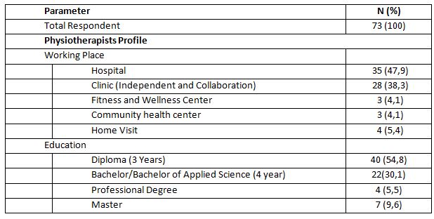
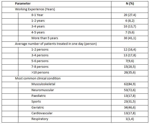
From 189 target respondents, only 73 respondents were willing to fill out the questionnaires given by the researchers. The physiotherapist’s identity is in the following table of respondent profiles.
Table 1. Profile of Respondent
It can be seen that of 73
physiotherapists as respondents in this survey, 35 people or around 47.9%
worked in hospitals and only 4 people or around 4.1% worked in community health
centers. The previous study also stated that in a hospital, there were more
than 10 physiotherapists8. The workplace of
physiotherapists in some countries is also dominated by hospitals and clinics10,11. In terms of
education level of the respondents, as many as 40 respondents or around 54.8% had
a diploma which is three-year study in university level.
There were 7 people or around 9.6% who
had a master’s degree. In Indonesia, those who are given the authority to
practice physiotherapy are physiotherapists starting at the diploma level to
the professional level2. This is already
in line with the provisions of the World Physiotherapy Organization, although
there is still a national government that gives authority to physiotherapists
at the diploma level1. Thus,
physiotherapists in East Jakarta are considered to meet the minimum
qualifications to practice physiotherapy. Likewise, in some countries, it is
found that the qualifications for physiotherapy education are quite varied and
still follow the national regulations of each country10,11.
The following result of the
questionnaire is about the physiotherapists’ work experience which describes
the range of work of physiotherapists, the average number of patients treated
in one day, and the condition of patients treated by the physiotherapists. The
data are presented in the following Table 2.
The following result of the questionnaire is about the physiotherapists’ work experience which describes the range of work of physiotherapists, the average number of patients treated in one day, and the condition of patients treated by the physiotherapists. The data are presented in the following Table 2.
Table 2. Patients Demographic
In the table, it can be seen that the
majority of respondents who have worked for more than five years are 41.1%,
which indicates that the physiotherapists have experienced in working and using
EPAmodalities. Physiotherapists with more than five years of experience will
indeed provide better patient satisfaction12.
As many as 35.6% physiotherapists
treated more than ten patients daily and around 16.4% treat one to two patients
every day. From these data, it can be seen that some physiotherapists still
treat too many patients in one day, that is, more than ten patients daily. If
there are more than 10 patients treated daily by the physiotherapist, assuming
one patient is an hour, it indicated that the physiotherapist in providing
services is not optimal, because in general, the number of daily working hours
is only about 8 hours11–13. However, there
are not many studies that support this. It can also cause harm to the
physiotherapist who is likely to experience fatigue. Thus, the number of
physiotherapists must be increased in order to provide optimal services14.
Most respondents or 84.9% treated
patients with musculoskeletal condition and the least treated were patients with
respiratory condition. Several studies also support this. As in a study by Jahan et al., (2021) who found that
physiotherapists mostly treated musculoskeletal patients. Likewise, in a study
in a hospital, physiotherapists generally dealt with various types of patients
with musculoskeletal disorders8. In addition, a
study in a certain region also stated that musculoskeletal cases were very
dominant in the physiotherapy practice15.
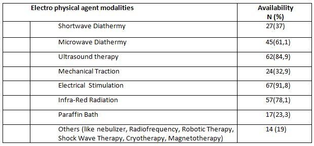
The following result is related to the availability of EPA modalities as well as the frequency of use. Table 3 shows the available modalities in each physiotherapist’s workplace.
Table 3. Availability of Electro Physical Agent Modality
In the view from the availability of
EPA modalities found in each respondent’s workplace, most respondents or 91.8%
answered the availability of electrical stimulation. It is followed by EPA modalities
in the form of ultrasound therapy (84.9%). The least EPA modalities found in
the respondent’s workplace were paraffin bath (23.3%). The availability of
diathermy modalities in the form of Micro Wave Diathermy at the respondents’
workplace had a considerablevalue of around 61.1%, including Short Wave
Diathermy which had a considerable percentage (37%).
There are also heat therapy modalities
that are quite popular among physiotherapists, namely Infrared Radiation device
which was answered by 50% of the total respondents. A study in Australia also
shows that ultrasound therapy is a modality that has considerable availability16. Furthermore, the
use of EPA interventions is still a trend among physiotherapists in Asia4,17.
However, apart from Asia, in America
there is also a trend in the use of EPA18. The most widely
available modality tools are cold and hot agents, with electrical stimulation
therapy and ultrasound therapy. It was also found that in America, there are
very few diathermy modalities available.
From the results of this study, it can
be seen that physiotherapists in East Jakarta have the availability of adequate
EPA modalities in accordance with the needs of the patients. Nevertheless, the
needs for EPA must really be acknowledged and the latest research developments
should keep up with the effectiveness of existing EPA modalities(Belanger, 2015; Bellew et al., 2016;
Goh &Abe, 2015). There are also
some EPA modalities in the table 3 that tend to be new and have very little
availability.In addition, the existing modalities data can also be a reference
for lecturers to continue to provide updates on the science of EPA modalities.
By understanding the availability, the lecturer needs to provide understanding
to physiotherapy students at the university in order to be more critical in the
use of electrophysical instruments in clinical practice. Previous studies also
used the survey results in the availability of physiotherapy modalities for
teaching purposes at universities18.
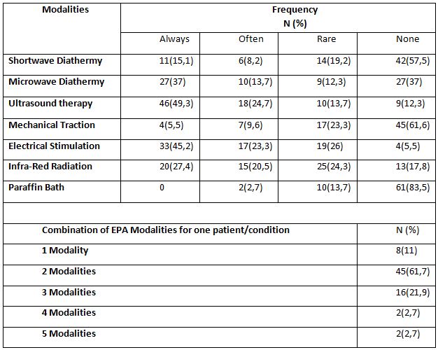
The last results of this study were
related to the frequency of the use of EPA. In this study, we only prepare the
types of modalities that are popularly used by physiotherapists, which are SWD,
MWD, US, IRR, Mechanical Traction, Electrical Stimulation, LASER, and paraffin
bath. It is also based on the availability of such modalities at the
physiotherapists’ place of practice. Moreover, we conducted a survey related to
how many combinations of EPA modalities were used by physiotherapist for each
patient with certain condition.
The time frame we chose was one week to make it easier for the physiotherapists to remember the modalities they used on average. Frequency classification was also made using the range: always (everyday), often (four to five times a week), rarely (one to three times a week), and none (not using the modality or the modality is not available). All usage and frequency data are shown in Table 4.
Table 4. Frequency of Electro Physical Agent Usage
From the results above, it can be seen that the use of EPA modalities in the form of ultrasound therapy and electrical stimulation was the most widely used. Subsequently, as many as 49.3% of respondents used the ultrasound therapy modality every day and only 12.3% never used the ultrasound therapy modality. A total of 45.2% of respondents used electrical stimulation modality and only 5.5% never used it. The modalities of paraffin bath and mechanical traction were the least used EPA modalities. As many as 83.5% of respondents never used paraffin bath and 61.6% of respondents never used mechanical traction.
The use of electrical
stimulation is believed to reduce neuromusculoskeletal disorders. As in the
results of a previous study by Manik &
Rahmansyah (2021), it mentioned that EPA modalities in the form of electrical
stimulation can reduce pain in neuromuscular and musculoskeletal conditions20. In line with this, a study by Abe
et.al (2016), which surveyed 1099
respondents from 170 hospitals/clinics showed that the use of electrical
stimulation in the form of low frequency currents and ultrasound therapy ranked
second and third below the modality in the form of hot pack4. Furthermore, a study by Greco et
al. (2018) also found that the use of electrical
stimulation was quite dominant, along with ultrasound therapy and thermal
modality (cold or hot). In addition, we should take attention to the results of
the existing research is that despite the availability of diathermy
physiotherapy tools, it does not guarantee that they are often used. The trend
that is developing in the world is that diathermy modality should be abandoned
and replaced with more effective modality 9,18,19.
In
this study, it was also found that physiotherapists used more than two
modalities in one patient with one condition. This shows that physiotherapists
very often used EPA modalities. The use of more than two modalities is possible
if the patient’s condition requires intensive intervention and there are more
than two symptoms in one condition19,21. Knowledge of the use of appropriate
and effective interventions needs to be carried out in the future to be able to
provide optimal services to the patients22,23.
This study is a very simple
survey as a preliminary study. More complex analysis in the existing data can
be done as a development of future research. Likewise, the survey in this study
was only conducted in one city, so more detailed research using a larger
population is needed as a development in the future. This study also has many
limitations, one of which is the willingness of the physiotherapist to fill out
the questionnaire. Therefore, it is possible to conduct more interesting
research that can attract the attention of the physiotherapist to fill out the
questionnaires in the future. The reasons for selecting interventions related
to the condition of the patients have not been captured in this study, so
further research development is needed.
CONCLUSION
The EPA modalities are
quite widely available among physiotherapists in East Jakarta, Indonesia, both
in health facilities and independent clinical practices. The most widely
available modalities are electrical stimulation, ultrasound therapy, and
infrared radiation and in addition to the diathermy modality, which is also
quite widely available. The availability of EPAtools is also in line with the
frequency of use dominated by ultrasound therapy, electrical stimulation, and
Microwave Diathermy. Physiotherapists also quite often use EPA because in one
patient with one condition, physiotherapists frequently use more than one
modalities.
Recommendation: Physiotherapists in East Jakarta should
pursue their education to a higher level to find out the latest developments in
electrophysical interventions. The use of diathermy should also be reduced by
its decreasing use abroad. The number of patients handled by one
physiotherapist in one day is too many, so it is necessary to add the number of
physiotherapists who work in a hospital. Based on the result, it also suggested
that at universities, it is still necessary to provide scientific development
in EPA, especially the frequent modalities like ultrasound therapytherapy,
electrical stimulation, and diathermy so that when the students graduate, they
can practice well.
Acknowledgement: The researcher would like to thank
every physiotherapist who is willing to participate in this research.
Furthermore, we also thank the of the Indonesian Physiotherapy Association,
East Jakarta Branch, for allowing us to collect data from the organization
members.
Conflict of interest: The author has no conflict of interest
to declare.
Funding of study: This study was funded by Universitas
Kristen Indonesia.
Compliance with Ethics: This research does not provide anything that is harmful to the
respondents so that it does not use ethical research. However, this research
was conducted with the permission of the university and the permissionfrom
chairman of the Indonesian physiotherapy association, East
Jakarta branch, with reference number 03/IFI-JAKTIM/XI/2021.
REFERENCES
1. World Confederation for Physical Therapy.
Guideline for standards of physical therapy practice. Published online
2011:1-19. https://world.physio/sites/default/files/2020-07/G-2011-Standards-practice.pdf
2. Kementerian Kesehatan Republik Indonesia. Peraturan Menteri Kesehatan No.80 Tahun 2013.;
2013.
3. Watson T. The role of electrotherapy in
contemporary physiotherapy practice. Man
Ther. 2000;5(3):132-141.
4. Abe Y, Goh AC, Miyoshi K. Availability,
usage, and factors affecting usage of electrophysical agents by physical
therapists: A regional cross-sectional survey. J Phys Ther Sci. 2016;28(11):3088-3094. doi:10.1589/jpts.28.3088
5. Starkey C. Therapeutic Modalities. Fourth Edi. (McDonald Q, ed.). F. A.
Davis Company; 2013. www.fadavis.com
7. Kementerian Kesehatan Republik Indonesia. Peraturan Menteri Kesehatan Republik
Indonesia Nomor 65 Tahun 2015 Tentang Standar Pelayanan Fisioterapi. Vol
16.; 2015.
8. Panjaitan LA. Penggunaan Terapi Elektrofisis
Pada Satu Rumah Sakit Umum Swasta di Jakarta. J Fisioter. 2020;20(2):40-45.
9. Goh A-C, Abe Y. New directions in
electrophysical agents : where do we go from here? Japanese J Electrophysical Agents VO – 22. 2015;(April):4.
10. Khairy WA, Bekhet AH, Sayed B, Elmetwally SE,
Elsayed AM, Jahan AM. Prevalence, profile, and response to work-related
musculoskeletal disorders among egyptian physiotherapists. Open Access Maced J Med Sci.
2019;7(10):1692-1699.
11. Nkhata L a, Zyaambo C, Nzala SH, Siziya S.
Work-related Musculoskeletal Disorders : prevalence , contributing factors and
coping strategies among Physiotherapy personnel in Lusaka , Kitwe and Ndola
districts , Zambia. Physiotherapy.
2010; 37(4):262-267.
12. Jahan AM, Rwaiha AE, Gusaibat SR, Al-Ahwal NA,
Al-Jafairi ZM, Al-Rashidi MA. Patient Satisfaction With Physiotherapy Services
in Libya: A Cross-Sectional Study. J
Patient Exp. 2021;8:1-7.
13. Hima Bindu P, Thiruppathi A. Work Related
Musculoskeletal Discomfort (WRMSD) among Physiotherapists. Int J Physiother. 2014;1(4):200.
14. Manurung NSA, Sunaryo T, Gunawan I, Anggiat L.
Analysis of the need for Physiotherapists in a private hospital in Indonesia
using the workload indicator of staffing need referring to the implementation
of the physiotherapy process as risk mitigation of services. Int J Med Exerc Sci.
2020;06(01):697-705.
15. Odumodu IJ, Olufunlayo TF, Ogunnowo BE, Kalu
ME. Satisfaction With Services Among Attendees of Physiotherapy Outpatient
Clinics in Tertiary Hospitals in Lagos State. J Patient Exp. 2020;7(4):468-478.
16. Chipchase LS, Williams MT, Robertson VJ. A
national study of the availability and use of electrophysical agents by
Australian physiotherapists. Physiother
Theory Pract. 2009;25(4):279-296.
17. Shah SGS, Farrow A. Trends in the availability
and usage of electrophysical agents in physiotherapy practices from 1990 to
2010: a review. Phys Ther Rev.
2012;17(4):207-226.
18. Greco JL, Lamberg EM, McKenna RF, Muratori LM.
Trends in availability and usage of biophysical agents among physical
therapists in the United States. Phys
Ther Rev. 2018;23(2):116-123.
19. Bellew JW, Michlovitz SL, Nolan TP. MODALITIES For Therapeutic Intervention.
F. A. Davis Company; 2016.
20. Manik JWH, Rahmansyah B. The effect of nerve
mobilization on the median nerve in pain perception of electrical stimulation. Int J Med Exerc Sci |2021;7(3). 2021;
7 (August):1104-1112.
21. Kim MK, Ji SG, Cha HK, Chang JS. Effects of
electromagnetic diathermy in conjunction with nerve mobilization in the
management of lower back pain. J Phys
Ther Sci. 2012;24(12):1337-1339.
22. Beales D, Mitchell T, Holthouse D. Stepped care
for musculoskeletal pain is ineffective: A model for utilisation of specialist
physiotherapists in primary healthcare management. Aust J Prim Health. 2021;27(6):431-436.
23. Tiktinsky R, Chen L, Narayan P. Electrotherapy: Yesterday, today and tomorrow. Haemophilia. 2010;16(SUPPL. 5):126-131.
Citation: Beriman Rahmansyah, Lucky Anggiat. Availability and usage of electro physical agentmodality by Physiotherapist in East Jakarta, International Journal of Medical and Exercise Science, March 2022; 8(1); 1228-1237.
Background: Diastasis Recti Abdominis (DRA) is a stretching and widening of the linea-alba which is a connective tissue that stretches in the middle of the abdomen that occurs in the second trimester to the third trimester and will continue until after delivery. Usually it cause complaints such as abdominal muscle weakness, lower back pain and posture disorders. Efforts that can be made to reduce the dilation of the postnatal linea-alba are by therapeutic exercise or physical exercise on the abdominal muscles. This study will focus on plank exercise was conducted to determine its effect on reducing the distance of the DRA below umbilicus.
Method: This research is a quantitative analysis with a quasi-experimental design that uses a two-group approach where the treatment was only given to one group and the other group only as a control.
Result: There was a reduction in the width of the diastasis rectus abdominis below the umbilicus by (67.7%) or as many as 42 people who did plank exercise while in the control group only (75.8%) or as many as 47 people who experienced a reduction in the width of the DRA lower umbilicus.
Conclusion: There is an effect of plank exercise on changes in the distance of the DRA below umbilicus and there is a relationship between plank exercise and a reduction in the width of the DRA below umbilicus in postpartum women.
Received on 4thFebruary 2022, Revised on 19thFebruary 2022, Accepted on 26thFebruary 2022, DOI:10.36678/IJMAES.2022.V08I01.007
INTRODUCTION
Diastasis Recti Abdominis (DRA) is stretching
and widening of the linea alba, which is the connective tissue that runs down
the middle of the abdomen and connects major abdominal muscles such as external
obliques, internal obliques, transversus abdominis, and rectus abdominis1. Some researcher also stated that this condition usually happens when entering the second trimester and will become clearer
in the third trimester and will continue after childbirth (postpartum)2,3.
Diastasis Recti Abdominis will widen due to
frequent pregnancies. It also occurs because during pregnancy there is an
increase in body weight and an increase in the hormonal levels of relaxin,
progesterone, and estrogen from the connective tissue which causes mechanical
pressure on the abdominal wall by the growing fetus so that the connective
tissue becomes soft and the linea alba becomes tenuous4.
Almost 100% of pregnant women experience DRA5,6. Approximately 50% of nulliparous women experienced DRA
and in women who undergo abdominal surgery and in postmenopausal patients7.Factors causing DRA are found in women who do excessive
abdominal exercises especially in the first trimester, women who like to use
hormone therapy, women who perform repeated operations on their abdomen, women
with multiple pregnancies, large babies, and caesarean sections8. Ambarwati and Candido also stated that age and
multiparity can be risk factors for rectus abdominis diastasis in women9,10.
Several studies have stated that the general
impacts of DRA are the weakness of the
abdominal muscles, urinary incontinence, decreased elasticity of the abdominal
wall, functional and cosmetic disorders, low back pain5,6,9. Furthermore, about 52% of patients with urogynecological
disorders stated that they had DRA and about 66% had complaints of pelvic floor
muscle weakness such as stress urinary incontinence, stool incontinence and/or
pelvic organ prolapse7.
Efforts that can be made to reduce the dilation
of the postnatal linea alba are by therapeutic exercise or physical exercise on
the abdominal muscles4. Previous research by Gitta et al. (2016) which stated that static contraction exercises in the abdominal muscles
have been shown to reduce the DRA distance11. Research by Acharry & Kutty (2015) also stated that
to prevent and reduce the DRA, physical therapy can be carried out after
childbirth12. Another research by Wijayanti, (2016) proved that abdominal circumference can be reduced by
strengthening abdominal muscles, sit-ups,
postpartum exercise, physiotherapy, and plank exercise13.
Plank exercise is a type of static contraction
exercise that is isometric exercise, which is
muscle contraction exercises against resistance without causing changes in
muscle length and joint motion14. This exercise can activate neuro-adaptive and proprioceptor
mechanisms through altered sensory input to muscles. Lee et al.,stated that
muscle activity in plank exercise is related to posture and muscle position
exercises and has been proven to strengthen core muscles (especially transverse
abdominis)15.
Based on all previous evidences obtained that
exercise and physical exercise of the abdominal muscles can prevent and reduce
postnatal women’s problems such as reducing abdominal circumference and
increasing abdominal muscle strength. This study willfocus on plank exercise
was conducted to determine its effect on reducing the distance of the DRA below
umbilicus.
METHODS
This research is a quantitative analysis with a
quasi-experimental design that uses a
two-group approach where the treatment was only given to one group and the
other group only as a control16. Meanwhile, the research design used pre and post-test design
and how to take samples using a purposive
sampling method.This study aims to determine the effect of plank exercise on DRAin women postpartum.
Before and after being given treatment, the sample was measured for the length
of the uterus by palpation and using a measuring device,calipers. The number of
postpartum women who participated in this study was 124 people and was
divided equally into 62 people in each group.
This research was conducted in the working area
of the Makassar District Health Center, East Jakarta, and was carried out from April 2019 to June 2019. Plank
Exercise intervention is carried out 3 times a week for 6 weeks.
The criteria set for the research sample are as
follows:
Inclusion
criteria:
All postpartum women at the Makassar District Health
Center, East Jakarta and the Kebon Pala Village Health Center, East Jakarta
Postpartum woman with minimum 2 hours after delivery
Postpartum women with DRA (>2.5 cm)
Postpartum women with a disease history of vaginal delivery
Willing to follow an exercise program regularly.
Willing to not do activities that are suspected of affectingDRA, such as participating in other gymnastics activities
outside the given program.
Willing to be a sample and sign informed consent.
Exclusion
Criteria:
Postpartum women who refused to participate in this study
Postpartum women who have participated in postpartum
gymnastics and other physical activities
Postpartum women who are having heart problems, respiratory
problems including excessive coughing or sneezing
Postpartum women with pelvic or abdominal surgery (except
for cesarean section), neurological disease, bowel or bladder trauma
Depressed postpartum woman
Measurement
of DRA:
Measurement by palpation of the fingers and
calipers DRA measurements were carried out before and after plank exercise.
According to Bursch, Mantle et al. and Rett et al. (2009), the palpation
measurement technique meets the criteria of reliability and ease to use. The
measurement can be done by placing a finger palpation between the medial border
of the right and left rectus abdominis muscles, which is parallel to the linea
alba3,17,18.
The size of DRA is determined by the number of fingers that enter between the two gaps in the abdominal wall that are the muscles above and below the umbilicus when the individual performs the movement placing her chin on her chest. Clinically, this palpation technique is simple.
Figure 1. The Measurement of DRA with Palpation technique 5
1.Measurement procedure by palpation is carried
out as follows:
Make the subject lying flat on her back without a pillow
under her head.
Place the fingertips of one hand on the midline of the
abdomen with the tip of the index finger just below the umbilicus and the other
finger lined longitudinally downward toward the symphysis pubis.
Ask the subject to raise her head to rest her chin on her
chest in the area between her breasts. Make sure not to press her hands against
the bed or grip the mat to help her, as this prevents the use of the abdominal
muscles.
As the subject attempts to rest her chin between her
breasts, gently press your fingertips close to her abdomen. You will feel the
abdominal muscles like two rubber bandages approaching the midline from either
side.
Measure the gap between them with calipers when the muscles are contracted.
Note the distance between the two slits
When lowering the head, the abdominal muscles move further
apart and are less distinguishable when the muscles are relaxed. Your
fingertips will follow the rectus muscle apart to the lateral side of each
abdomen.
Measure the distance between the two rectus muscles when
they are relaxed.
Note the measurement results
2. Measurement using calipers
Measurement procedure the rectal distance was
measured using a caliper technique, the participant was in a lying position
with a pillow under the head and feet resting on the base and arms by the side
of the body. The desired measurement location is marked 4.5 cm above the
midpoint of the umbilicus and 4.5 cm below the midpoint of the umbilicus. Each
participant raises their head until the spine of the scapula is off the table
surface.
Participants were asked to maintain this position while the examiner palpated the medial border of the right & left abdominis abdominal muscles at the marked area. Measuring the inside of the jaws of a digital nylon caliper, positioned at the location of the palpated finger, perpendicular to the direction of the muscle and adjusted for the width of the perceived intra-rectal distance. Three measurements were taken for each assessment and then recorded. Participants are allowed to rest between measurements and are also allowed to rest if they feel tired 21.
Figure 2. Measuring the interrecti distance 4.5 cm above and below the umbilicus 19
Intervention Group
In the treatment group, interventions were given in the form of plank
exercise.Plank exercise is an isometric type of exercise, which is a type of
static muscle contraction exercise against resistance without any change in
muscle length or not followed by joint movement14. This exercise begins by extending
the legs while supporting the upper body with the arms. Bend elbow position,
hold abdominal and gluteal muscles.
This exercise is done 3 times a week for 6 weeks with 3 repetitions of each exercise. The prone plank exercise is carried out by holding it for 30 seconds for each movement then resting for 15 seconds and then repeating it 20.
Figure 3. Plank Exercise 20
Control
Group
In the control group, the research sample was
given an education program in the form of counseling on the importance of
exercise to restore the health of postpartum women with DRA conditions. Based on research, regular exercise can also improve the DRA4.
RESULTS AND DISCUSSION
Following is the distribution of Plank Exercisevariables and the control group
Table 1. Frequency Distribution of Independent Variable
Based on table 1, each postpartum woman performs Plank Exercise that the intervention group also included 62 people (50%) and the control group with 62 people (50%).
Table 2. Frequency Distribution of Confounding Variables
Based on table 2, the number of postpartum women
aged < 20 years or > 30 years was 59 people (47.6%), multiparous status
was 59 people (47.6%), had Obesity/Overweight BMI which was 60 people (48.4%),
history of singleton pregnancy namely 120 people (96.8%), and the weight of
newborns was normal (not macrosomic) that was 112 people (90.3%).
Relationship
between Independent and Confounding Variables on Reduction of the width of the
DRA below umbilicus)
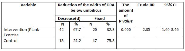
Table 3. shows that those who did plank exercise
mostly reduced the width of the DRA below umbilicus was 42 people (67.7%).
While
most of the postpartum women who do not do
plank exercise did not experience a reduction in the width of the DRA below
umbilicus (fixed) was 47 people (75.8%). Chi
Square test results stated that there is a relationship between plank exercise
with a reduction in the width of the DRA below umbilicus (p value <0.05).
Postpartum women who did Plank Exercise had a 2.3 times chance of experiencing a reduction in the width of the DRA below umbilicus compared to those who did not do it (p value =0.000; cRR=2.35;95%CI= 1.60-3.46).
Table 3. Relationship of Plank Exercise with Reduction of the width of DRA below umbilicus
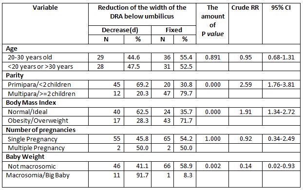
Based on Table 4, the results of the cross-tabulation show that most of the postpartum women aged < 20 years or >30 years did not experience a reduction in the width of the DRA below umbilicus (fixed) was 31 people (52.5%). While those aged 20-30 years most did not experience a reduction in the width of the DRA below umbilicus (fixed) was 36 people (55.4%). Chi Square test results showed that there was no relationship between age and a reduction in the width of the DRA below umbilicus (p value> 0.05).
Table 4. Relationship of Confounding Variables with Reduction of the DRA below umbilicus
The results of cross tabulation showed that most
of the postpartum women with multiparous status did not experience a reduction
in the width of the DRA below umbilicus (fixed) was 47 people (79.7%). While most of the primipara experienced
a reduction in the width of the DRA below umbilicus was 45 people (69.2%). Chi Square test results stated that
there is a relationship between plank exercise with a reduction in the width of
the DRA below umbilicus (p value <0.05). Primipara postpartum women were 2.6
times more likely to experience a reduction in the width of the DRA below
umbilicus than multiparous women (p value
=0.000; cRR=2.59;95%CI= 1.76-3.81).
The results of the cross tabulation showed that
the postpartum women who were obese/overweight
mostly did not experience a reduction in the width of the DRA below umbilicus
(fixed) was 43 people (71.7%). Meanwhile, postpartum women with normal/ideal BMI experienced a reduction in the
width of the DRA below umbilicus was 40 people
(62.5%). Chi Square test results showed that there was a relationship between
excess BMI and a reduction in the width of the DRA below umbilicus (p value
<0.05). Postpartum women with normal/ideal BMI were 1.9 times more likely to
experience a reduction in the width of the DRA below umbilicus compared to
obesity/overweight (p value=0.000; cRR=1.91;95%CI=1.34-2.72).
The results of cross tabulation showed that
postpartum women with multiple pregnancies 50% experienced a reduction in the
width of the DRA below the umbilicus. While postpartum women with singleton pregnancies mostly did not experience
a reduction in the width of the DRA below the umbilicus (fixed) was 65 people (54.2%). Fisher Exact test results showed that
there was no relationship between multiple pregnancies with a reduction in the
width of the DRA below umbilicus (p value> 0.05).
The results of cross tabulation showed that postpartum women who gave birth to babies weighing 4000 grams mostly
experienced a reduction in the width of the DRA below the umbilicus was 11 people (91.7%). Meanwhile, postpartum women with babies weighing <4000 grams mostly did not
experience a reduction in the width of the DRA below umbilicus (fixed) was 66 people (58.9%). Chi Square test results showed that
there was a relationship between large baby weight and a reduction in the width
of the DRA below umbilicus (p value <0.05). Postpartum women with not
macrosomic babies were 0.1 times more likely to experience a reduction in the
width of the DRA below umbilicus compared to women with macrosomic babies (p
value=0.002; CRR=0.14;95%CI=0.02-0.93).
The results of this study are in line with
research by Fitriahadi 21. With a quasi-experimental research model and similar
types of interventions. The results of the study stated that most of the
decrease in DRA occurred quickly in the treatment group (plank exercise) by 10
(66.65), while a slower decline in DRA occurred in the untreated group (control
group) by 9 (60%), supported by bivariate analysis with p value = 0.003, this
proves that there was an effect on strengthening the rectus abdominis muscle to
reduce the DRA distance in postpartum women.
The results of the study are in line with
previous research that there was an effect of sit-up exercise with prone plank
exercise on decreasing abdominal circumference in adolescent girls13. Other research also stated that exercise will have an
effect after being done for 6 weeks, for example, weight training can increase
muscle strength by 20.1% within those weeks22. In general, previous research by Khandale & Hande
also stated that abdominal muscle training can reduce DRA in early postpartum
women and can prevent complications due to DRA23.
This research still has some limitations. This
research method is still very simple and needs to be improved for the better in
future research. The examination is also carried out in a simple manner, the
use of other, more sophisticated examination tools is needed in future
research. Likewise, this study only evaluates short-term effects, then,
long-term research to determine long-term effects also needs to be done. The
use of the results of this study must be responsible to avoid misunderstanding.
CONCLUSION
Based on data analysis, it was found that there
was a reduction in the width of the DRA below
umbilicus in postpartum women with treatment of 42 people (67.7%) while in the
control group only 15 people (24.2%). Thus, it can be concluded thatthere is an
effect of plank exercise on changes in the distance of the DRA below umbilicus
and there is a relationship between plank exercise and a reduction in the width
of the DRA below umbilicus in postpartum women.
Recommendations:
As a recommendation from this study,
physiotherapists would be able to advise postpartum women to do plank exercise
in reducing various postnatal complaints and increasing quality of life both in
the community and at home.
Ethical
Clearance: This research was approved by
the East Jakarta Health Service Center with reference number 1950/1.772.2.
Acknowledgements:
We would like to thank the
postpartum women in Makassar Health Center, East
Jakarta who are willing to participate in this research.
REFERENCE
1. Lee D, Lee L, McLaughlin L. Stability, continence and breathing: The role of fascia following pregnancy and delivery. J Bodyw Mov Ther. 2008;12(4); 333-348.
2. Estiani
M, Aisyah A. Faktor-Faktor Yang Berhubungan Dengan Kejadian Diastasis Rekti
Abdominis Pada Ibu Post Partum Di Wilayah Kerja Uptd Puskesmas Sukaraya
Baturaja. J Keperawatan Sriwij.
2018; 5(2); 24-31.
3. Rett
MT, Braga MD, Bernardes NO, Andrade SC. Prevalence of diastasis of the rectus
abdominis muscles immediately postpartum: Comparison between primiparae and
multiparae. Brazilian J Phys Ther.
2009; 13(4) ;275-280.
4. Michalska
A, Rokita W, Wolder D, Pogorzelska J, Kaczmarczyk K. Diastasis recti abdominis
– A review of treatment methods. Ginekol
Pol. 2018; 89(2):97-101.
5. Mota
P, Pascoal AG, Carita AI, Bø K. Normal width of the inter-recti distance in
pregnant and postpartum primiparous women. Musculoskelet Sci Pract. 2018;35; 34-37.
6. Walton
LM, Costa A, LaVanture D, McIlrath S, Stebbins B. The effects of a 6 week
dynamic core stability plank exercise program compared to a traditional supine
core stability strengthening program on diastasis recti abdominis closure,
pain, oswestry disability index (ODI) and pelvic floor disability index score. Phys Ther Rehabil. 2016;3(1); 3.
7. Spitznagle
TM, Leong FC, Van Dillen LR. Prevalence of diastasis recti abdominis in a
urogynecological patient population. Int
Urogynecol J. 2007;18(3); 321-328.
8. Aswini
D, Srihari SK. An Overview of the Studies on Diastasis Recti Abdominis in
Postpartum Women. J Gynecol Womens
Heal. 2019; 14(5).
10. Candido
G. LT. JPA. Risk factors for diastatis of the recti abdominis. J Assoc Chart Physiother Women’s Heal.
2005;97(January 2005); 49-54.
11. Gitta
S, Magyar Z, Tardi P, et al. How to Treat Diastasis Recti Abdominis with
Physical Therapy: A Case Report. J Dis.
2016;3(2); 16-20.
12. Acharry
N, Kutty RK. Abdominal Exercise With Bracing, a Therapeutic Efficacy in
Reducing Diastasis-Recti Among Postpartal Females. Int J Physiother Res. 2015; 3(2); 999-1005.
13. Wijayanti
D. Perbedaan Pengaruh Sit-up Exercise Dan Prone Plank Exercise Terhadap
Penurunan Lingkar Perut Remaja Putri. Publ
Manuscript, Univ Aisyiyah Yogyakarta. Published online 2016; 1-16.
14. Schoenfeld
BJ, Contreras BM. The long-lever posterior-tilt plank. Strength Cond J. 2013; 35(3):98-99.
15. Lee
J, Jeong K, Lee H, et al. Comparison of three different surface plank exercises
on core muscle activity. Phys Ther
Rehabil Sci. 2016;5(1); 29-33.
16. Sugiyono.
Metode Penelitian Pendidikan
Pendekatan Kuantitatif, Kualitatif Dan R&D. Alfabeta; 2013.
18. Mantle
J, Haslam J, Barton S. Physiotherapy
in Obstetrics and Gynaecology. Elsevier Ltd; 2004.
19. Chiarello
CM, McAuley JA. Concurrent validity of calipers and ultrasound imaging to
measure interrecti distance. J Orthop
Sports Phys Ther. 2013;43(7); 495-503.
20. Bennett
VR, Brown LK. Myles Textbook for
Midwives. Churchill Livingstone; 1999.
21. Fitriahadi
E. Pengaruh Penguatan Otot Rectus Abdominis Terhadap Penurunan Tfu Pada Ibu
Postpartum Pervaginam Di Bpm Kabupaten Sleman. J Kebidanan. 2019; 8(1); 61.
22. Sudarsono
S. Penyusunan Program Pelatihan Berbeban Untuk Meningkatkan Kekuatan. J Ilm SPIRIT. 2015;12(1); 31-43.
23. Khandale SR, Hande D. Effects of Abdominal Exercises on Reduction of Diastasis Recti in Postnatal Women. Int J Heal Sci Res. 2016; 6(6); 182. www.ijhsr.org
Citation: Lisnaini. Effect of plank exercise on diastasis recti abdominis lower umbilicus in postpartum women, International Journal of Medical and Exercise Science, March 2022; 8(1): 1219-1227.